
If you’ve ever had something caught in your eye, you know how irritating it is. If that something has ever scratched your eye (specifically your cornea), you know that initial irritation rapidly turns into pain. Dogs and cats have corneas too, and just like for you and me, they can be damaged by foreign particles. This week I share some important information about corneal ulcers and erosions in dogs and cats. Happy Reading!
Corneal Ulcers & Erosions – What are they?
The clear front part part of the eye is called the cornea, and it has five layers:
- Precorneal team film
- Epithelium and its basement membrane
- Stroma
- Descemet’s membrane
- Endothelium
The normal layers of the cornea from superficial to deep.
Corneal erosions occur when the epithelium is partially damaged. Corneal ulcers result from full-thickness damage to the epithelium that extends into the stroma. When an injury fully extends through the entire epithelium and stroma to Descemet’s membrane, the wound is called a descemetocele.
Corneal Ulcers & Erosions – What causes them?
Any number of problems may result in corneal erosions and ulcers. The most frequent cause is trauma to the cornea. Common traumatic injuries to the cornea include scratches (e.g: irritated cat swatting at impetuous dog), lacerations from sharp objects, prolonged exposure of the cornea due to incomplete of the eyelids (called lagophthalmos), and eyelash disorders (e.g.: distichiasis, trichiasis, ectopic cilia). Other causes of corneal erosions and ulcers are:
- Chemical burns
- Infections (e.g.: bacterial, viral, fungal)
- Other eye diseases (e.g.: keratoconjunctivitis sicca/dry eye, epithelial dystrophy)
- Diseases that start elsewhere in the body (e.g.: hypothyroidism, diabetes mellitus in dogs and cats, hyperadrenocorticism/Cushing’s disease)
Corneal Ulcers & Erosions – How are they diagnosed?
As a pet parent, you will likely seem some changes in your fur baby’s eye that raises concern. Clinical signs often associated with corneal ulcers and erosions include:
- Epiphora (excessive tearing)
- Reddening of the conjunctiva (the thin membrane over the sclera/”white of the eye”)
- Blepharospasm (involuntary closure of the eye)
- Photophobia (sensitivity to light)
- Change in color to the cornea (i.e.: may appear bluish/grey rather than clear)

A cat with blephoraspasm and epiphora of the left eye.
Ulcers and erosions typically are unilateral lesions, meaning only one eye is affected. They are typically not visible to the naked eye. Adequate visualization requires the use of special equipment and stain. Specifically, a veterinarian will place a drop of fluorescein stain, an orange-colored stain, that adheres to an eroded/ulcerated area and turns green. A special blue light and magnifying lens are then used to see the injury.
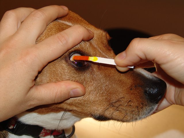
A veterinarian places a drop of fluorescein dye onto the cornea of a dog to highlight a corneal ulcer.

A corneal ulcer stained with fluorescein dye appears green when visualized with a special blue light source.
Corneal Ulcers & Erosions – How are they treated?
The treatment for an erosion or ulcer depends on depth of the injury. Superficial injuries are frequently treated with a topical antibiotic (to prevent a secondary bacterial infection) and a topical pain medication (to reduce spasms). This type of injury generally heals within one week.
Deeper injuries, including descemetoceles, may respond to eye medications, including ophthalmic pain and antibiotic solutions. The affected eye must also be protected from further injury. This may be done with an eye patch, Elizabethan collar, or a minor surgical procedure (to cover the ulcer or temporarily close the eyelids). For patients with deep corneal injuries, especially descemetoceles, consultation with a board-certified veterinary ophthalmologist is instrumental for maximizing the likelihood of a positive outcome.

The take-away message about corneal ulcers & erosions in dogs and cats…
Corneal ulcers and erosions are relatively common eye injuries in dogs and cats. Patients often have excessive tearing, reddening, and involuntary winking of the affected eye(s). A non-invasive test with a special stain is used to diagnose ulcers and erosions. The type of treatment depends on the depth of corneal injury. Partnering with a board-certified veterinary ophthalmologist can be helpful to help ensure a pet’s vision isn’t compromised.
To find a board-certified veterinary ophthalmologist, please visit the American College of Veterinary Ophthalmologists.
Wishing you wet-nosed kisses,
cgb