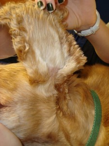
One of the most common ear problems in dogs is inflammation and/or infection of the outer ear; this is called otitis externa. This week I discuss some key features of this ailment with a sincere hope you will find the information helpful and share worthy. Happy reading!
Otitis Externa – What is the normal anatomy of the ear?
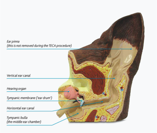
A dog’s ear has three major sections:
- The outer ear: This area of the ear consists of the ear flap (called a pinna) that funnels sound into the ear canal, a narrow L-shaped tube that travels to the deeper parts of the ear. The L-shaped canal is often referred to as having a vertical section and a horizontal one.
- The middle ear: The middle ear starts where the outer ear ends – at the level of the eardrum (called the tympanic membrane), a fragile membrane that can be easily damaged by disease. The middle ear is made of three small bones (called ossicles), an air-filled cavity (called a bulla) and a tube that leads from the bulla to the mouth (called the auditory or Eustachian tube).
- The inner ear: The inner ear connects to the brain and contains nerves that help with hearing and balance.
Otitis Externa – What causes it?
Board-certified veterinary dermatologists often categorize the causes of otitis externa in the three Ps:
Predisposing factors: These are factors that make a patient more susceptible to outer ear inflammation and infection. The narrow L-shaped anatomy of the ear canal allows for accumulation of secretions deep inside it. With swelling from inflammation and infection, narrowing of the canal is augmented and secretions can’t escape. Furthermore certain breeds, including Pugs, English Bulldogs, and Shar Peis, have a twist in the external ear canal that can completely occlude this tube when marked inflammation is present. Other predisposing factors including swimming, humidity, elevated ambient temperature, and trauma to the ear canal caused by inappropriate hair plucking and overly aggressive cleaning.
Primary factors: These are factors that directly cause inflammation in the outer ear, and include:
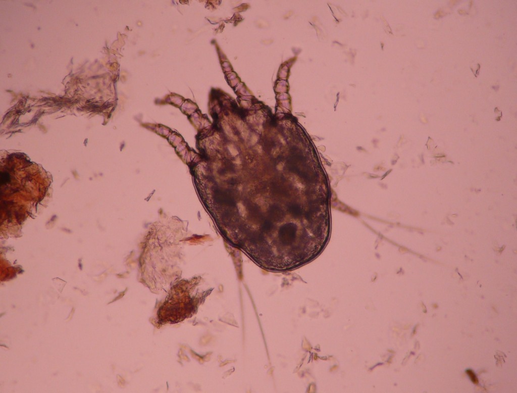
- Ear mites
- Ticks
- Food allergies
- Foreign objects (i.e.: grass awns)
- Polyps
- Tumors
- Hormone disorders
- Flea allergies
- Environmental allergies (called atopic dermatitis or atopy)
- Immune-mediated diseases (i.e.: pemphigus foliaceus)
- Certain genetic/hereditary disorders (i.e.: keratinization disorders)
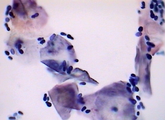
Perpetuating factors: These factors include infections with bacteria and yeast/fungal organisms.
Concurrent middle ear disease can also promote external ear inflammation and infection. Additionally chronic changes to ear anatomy may perpetuate ongoing inflammation, making infections more challenging to cure.
Otitis Externa – How is it diagnosed?
A veterinarian will employ a 3-pronged approach to the diagnosis of otitis externa:
- Obtaining a thorough patient history
- Performing a complete physical examination, including comprehensive ear and skin evaluations
- Using microscopy to look at cells and debris from the ear canal (called cytology)
Always be sure to provide very specific answers to questions asked by your pet’s veterinarian. Your responses could truly and directly impact the doctor’s diagnostic plan for your pet. A veterinarian will need to know if this is a first time issue (called acute otitis externa), if it’s one that initially improved but then recurred (called recurrent otitis externa), or if it’s one that has been going on for a while without resolution (called chronic otitis externa). Other important questions to answer for your pet’s veterinarian include:
- When did the clinical signs first occur?
- Has your dog ever had issues with excessive licking, scratching, chewing, etc.?
- Describe the environment in which your dog lives?
- Do any people in your dog’s residence have skin problems?
- Does your dog go to a boarding center, obedience school, and/or grooming facility?
- What is your dog’s diet?
A complete physical examination is uniquely important, as the concurrent presence of skin lesions and/or balance deficits helps a veterinarian develop a proper diagnostic plan. Evaluation of the external ear canal (called an otoscopic examination) is absolutely essential to the proper diagnosis of otitis externa. Below is a clip of a veterinarian performing video otoscopy in a dog.
A veterinarian will note the degree of inflammation and debris in the ear canal, as well as the presence of ulcers and the status of the eardrum (called the tympanic membrane). Understandably ears can be quite painful when inflamed and infected, and thus proper examination often requires sedation. If ear canals are swollen shut, temporary treatment with an anti-inflammatory medication may be needed to facilitate a complete ear evaluation.
Otitis Externa – How is it treated?
In general, treatment of otitis externa is directed at addressing both primary and perpetuating factors. Understandably the therapeutic approach can be quite variable given the myriad of factors that influence this disease. Cleaning ears is a critical part of every affected pet’s treatment plan, and a veterinarian or licensed veterinary technician should perform the initial cleaning. Of course continued intermittent cleaning at home will likely be recommended and necessary. Therefore families need to ensure they are comfortable performing such a task by spending adequate time with their pet’s healthcare team learning how to accomplish this cleaning effectively. Below is a video demonstrating the proper method for cleaning a dog’s ears.
Cleaning agents contain substances that soften and liquefy fat and wax in ear canal, making them easier to remove from this location. Proper cleaning also helps get rid of a substance bacteria make (called a biofilm) that reduces the efficacy of topical antibiotics. There are many cleaners available, and your veterinarian will make specific recommendations based on your pet’s specific condition and a family’s ability to perform proper at-home cleanings. For patients for whom at-home cleaning is not possible, your veterinarian may have some alternatives and/or may refer you to a board-certified veterinary dermatologist for further evaluation.
Veterinarians will tailor a pet’s medication regime based on history, physical examination, and ear cytology. Essentially all dogs with otitis externa benefit from a topical anti-inflammatory medication (most commonly a steroid); a veterinarian will make a decision about the use of topical and systemic antibacterial and/or antifungal medications based on ear cytology, severity of ear signs, and the overall health of the affected pet.
Patients with recurrent and chronic otitis externa require special attention, and consultation with a board-certified veterinary dermatologist is often uniquely beneficial. A thorough investigation for an underlying cause (i.e.: allergies, hyperadrenocorticism/Cushing’s disease) is absolutely essential and should not be ignored! Too often pets are prescribed treatment after treatment without a family allowing a veterinarian to make an adequate effort to address predisposing factors and identify primary and perpetuating ones. Partnering with a board-certified veterinary dermatologist can be a proverbial game-changer for these patients.
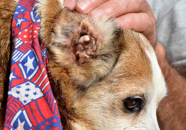
For some dogs, recurrent and chronic inflammation and infection damages the outer ear too badly. Resolution of outer ear disease even with proper cleaning and systemic and topical medications is simply not possible. Such patients need surgery to remove the ear canal (called a total ear canal ablation or TECA); this is a specialized surgery that should be performed by a board-certified veterinary surgeon.
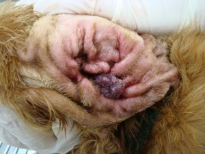
An ear with end-stage inflammation. This dog needs a total ear canal ablation (TECA) because systemic and topical medicines will not resolve this disease.
The take-away message about otitis externa…
Dogs are prone to outer ear infections because of the unique anatomy of their ears. Swelling and inflammation causes marked narrowing of the ear canal, contributing to the retention of moisture and secretions. A proper examination with a thorough diagnostic investigation is absolutely vital for developing the most effective treatment plan for affected pets. All patients with otitis externa benefit from anti-inflammatory steroid therapy and proper ear cleaning, the latter of which is essential before starting antibiotic or antifungal therapies. If too much damage has been done to the outer ear, surgery to remove the ear canal can restore patient comfort and provide permanent resolution. Consultation with a board-certified veterinary dermatologist can be invaluable for navigating the often frustrating waters of recurrent and chronic outer ear infections.
To find a board-certified veterinary dermatologist, please visit the American College of Veterinary Dermatology.
To find a board-certified veterinary surgeon, please visit the American College of Veterinary Surgeons.
Wishing you wet-nosed kisses,
cgb

