A common problem affecting the ears of dogs and cats is an aural hematoma. The condition is the collection of blood inside an ear flap, and can be very irritating to an affected pet. This week I share some tidbits about this problem, and I hope you find the information shareworthy. Happy reading!
Aural Hematomas – What causes them?
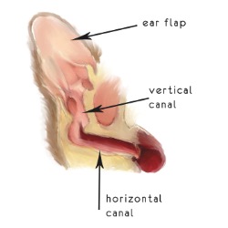
To understand this condition, one needs to have a basic understanding of the anatomy of a dog’s or cat’s ear flap (called a pinna). A pinna is composed essentially of skin and cartilage.
For reasons not yet completely understood, blood accumulates between the cartilage and the skin of a pinna. A common theory to explain the formation of aural hematomas is excessive head shaking induces small cracks/fractures in the cartilage and concurrently rupture small blood vessels. Blood subsequently pools between the skin and cartilage. A common reason for excessive head shaking in dogs in inflammation and infection of the external ear canal – this condition is called otitis externa. However, many pets who develop aural hematomas do not have a history of this condition.
Aural Hematomas – How are they diagnosed?
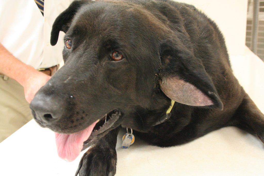
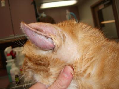
Diagnosing ear hematomas is relatively straightforward and is based on physical examination findings. They are documented more commonly in dogs than cats. Other potential causes of ear flap swelling are cysts, abscesses, and cancers. As mentioned earlier, many affected pets have a history of head shaking secondary to otitis externa. An ear with a relatively fresh aural hematoma looks like a major swelling of the ear flap. The enlargement is soft and fluid-filled. As days pass without intervention, the fluid becomes organized and scar tissue forms. Ear with chronic hematomas feel firm and the pinnae often appear disfigured. A veterinarian will evaluate the external ear canal for evidence of inflammation and infection.
Aural Hematomas – How are they treated?
Ear hematomas should be treated as quickly as possible. Both non-surgical and surgical techniques have been used successfully. A common treatment is non-surgical drainage of the fluid from the ear flap followed by instillation of a corticosteroid. There are several potentially successful surgical options, and most primary care veterinarians are both capable and comfortable surgically treating aural hematomas. For complicated and/or recurrent cases, it may be beneficial to consult with a board-certified veterinary surgeon or dermatologist.
The take-away message about aural hematomas in dogs & cats…
An aural hematoma, the accumulation of blood in the ear flap, is a common ear problem in dogs and cats. Veterinarians are able to readily diagnose this condition based on physical examination findings. Both non-surgical and surgical techniques have been used successfully to treat the condition.
To find a board-certified veterinary surgeon, please visit the American College of Veterinary Surgeons.
To find a board-certified veterinary dermatologist, please visit the American College of Veterinary Dermatology.
Wishing you wet-nosed kisses,
cgb


