One of the more common reasons patients are presented to me is for evaluation of an new and/or unusual lump or bump under the jaw. Arguably, the most common cause is an enlarged lymph node, but occasionally the problem is an abnormal salivary gland. This week’s post is dedicated to a salivary gland problem called a sialocele, and I hope you find the information interesting. Happy reading!

Sialoceles – What are they?
To appreciate sialoceles – also known as salivary mucoceles – one needs to have a basic understanding of salivary gland anatomy. Cats and dogs have four pairs of major salivary glands:
- Parotid salivary glands
- Mandibular salivary glands
- Sublingual salivary glands
- Zygomatic salivary glands

Salivary glands produce saliva that keeps the mouth, throat, and esophagus moist to facilitate delivery of food to the stomach. As saliva contains a variety of digestive enzymes, it also aids in the digestion of carbohydrates. A sialocele develops when a salivary gland or its duct is damaged, allowing saliva to inappropriately accumulate. Common causes of damage to salivary glands are:
- Trauma
- Cancer
- Obstruction of an associated salivary duct
- Foreign objects (i.e.: plant awns, splinters, etc.)
- Idiopathic (no identifiable cause)
What do sialoceles look like?
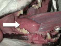
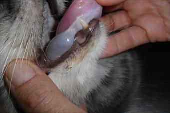
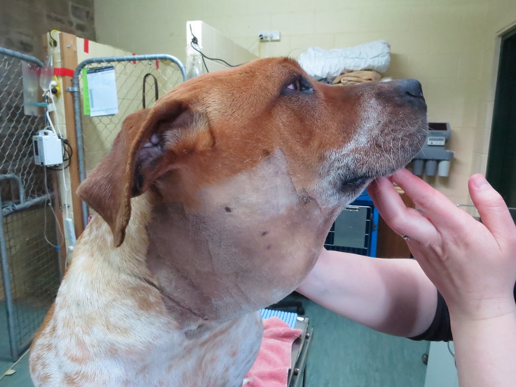
Sialoceles are most often recognized as soft, fluctuant, and non-painful swellings in the region of the affected salivary gland. For example, you may see a swelling under the base of the ear if your pet has sialocele affecting a parotid salivary gland. When the zygomatic salivary glands are involved, animals may have bulging of the eye or the roof of the mouth. A sialocele affecting a sublingual salivary glands is also called a ranula and often causes:
- Swelling under the tongue
- Abnormal tongue movement
- Oral bleeding
- Difficulty prehending food
- Difficulty swallowing
The sublingual glands are most commonly affected.


Sialoceles are typically classified by their location:
- Cervical (neck)
- Pharyngeal (throat)
- Sublingual (under the tongue)
How are they diagnosed?
A finding of a soft non-painful swelling in the region of a salivary gland is a major proverbial red flag for a salivary mucocele. A veterinarian will likely perform a fine needle aspiration (FNA) or fenestration of the swelling, a minimally invasive procedure using a vaccine-sized needle to aspirate fluid from the bulge. The fluid may be clear, yellowish, or blood-tinged, and special stains (i.e.: periodic acid Schiff) can be applied to confirm the presence of saliva.

How are they treated?
The treatment of choice for sialoceles is surgery. During surgery, the affected salivary gland is completely excised – this procedure is called a sialectomy. When cervical mucoceles are present, both the mandibular and sublingual glands need to be removed because they share a duct and capsule. The prognosis following surgery is very good!
The take-away message about sialoceles in cats & dogs…
The most common disease affecting salivary glands is a sialocele, an abnormal accumulation of saliva that is the result of damage to a salivary gland. Diagnosis is relatively straightforward, and salivary mucoceles may be effectively treated with surgery.
To find a board-certified veterinary surgeon, please visit the American College of Veterinary Surgeons.
Wishing you wet-nosed kisses,
CriticalCareDVM


