In previous posts I’ve written about a variety of heart diseases, including dilated cardiomyopathy in dogs, congestive heart failure, and heartworm disease. This week’s post is dedicated to another cardiac problem – hypertrophic cardiomyopathy (HCM) in cats. I hope you find the information helpful and shareworthy. Happy reading!
What is hypertrophic cardiomyopathy?
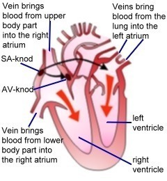
The heart is divided into a right side and a left side. Each side has one atrium and one ventricle. The right side of the heart receives de-oxygenated blood from the body; this blood first enters the right atrium, and the passes into the right ventricle where it is subsequently circulated through pulmonary arteries to the lungs to be oxygenated. The newly-oxygenated blood returns to the left side of the heart via pulmonary veins that deposit blood into the left atrium and then into the left ventricle. From the left ventricle blood is pumped into the aorta where it is ultimately distributed to all of the body’s tissues.
Hypertrophic cardiomyopathy is the most common primary myocardial (heart muscle) disease in cats, representing approximately 60% of heart diseases diagnosed in this species. The hallmark feature of HCM is thickening of the left ventricular wall. Specifically, the thickening is called concentric hypertrophy, and there’s no evidence the heart has to use more pressure to expel blood from the left ventricle.
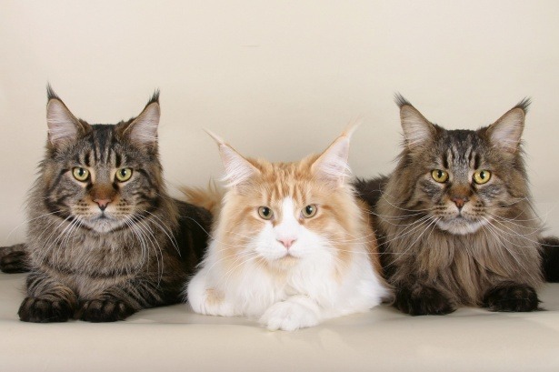
We don’t currently know the cause of HCM, but genetic abnormalities have been identified in Maine coons and ragdolls. In these breeds, two different genetic mutations in the cardiac myosin binding protein C gene have been documented, specifically A31P in Main coons and R820W in ragdolls. Several other breeds have been screened for mutations, but no others have been found to date.
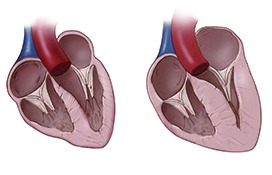
Heart muscle cells in HCM cats are not able to contract and relax normally, so they develop concentric hypertrophy of the left ventricle. As the disease progresses, heart muscle cells die and are replaced by fibrous (scar) tissue. These changes make muscle stiffer and less compliant. The cumulative alterations induced by scar tissue increase the pressure inside the left ventricle that dramatically affects blood flow through the heart. When the left ventricular wall thickens, the mitral valve moves abnormally, movement called systolic anterior motion (SAM) that produces a systolic heart murmur and further increases left ventricular pressure.
Pressure in the left atrium goes up because of left ventricular dysfunction, causing the chamber to markedly enlarge. When the left atrium enlarges, blood flow becomes more static. This predisposes cats to thrombus or abnormal clot formation. These clots can remain in the left atrium or can dislodge and embolize (get stuck in) the aorta, causing a life-threatening condition called feline arterial thromboembolism or FATE. Severely affected cats develop congestive heart failure (CHF).
What does it look like?
Across all cat breeds, males cats are overrepresented. However, male and female Main coons are affected equally. Hypertrophic cardiomyopathy is not present at birth, and HCM often doesn’t develop until middle age or old age. The exception is ragdolls who can develop HCM at less than one year of age.
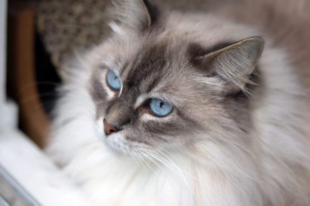
Most cats with HCM have no direct clinical signs of left ventricular dysfunction. Rather, cats are most often presented to their primary care doctor for clinical signs consistent with major sequelae of left ventricular dysfunction: congestive heart failure (i.e.: acutely increased respiratory rate & effort) and FATE. Clinical signs of FATE are often referred to as the 5 Ps:
- Pain
- Paralysis
- Pulses absent in affected limb
- Poikilothermia (cool affected limb)
- Pallor of the foot pads
How is hypertrophic cardiomyopathy diagnosed?
A veterinarian will thoroughly review a cat’s medical history and perform a complete physical examination. Cats with mild to moderate HCM often do not have clinical signs. Less than 50% of affected cats have a heart murmur, and only ~33% with a murmur have any form of cardiomyopathy.
According to board-certified veterinary cardiologist Dr. Kacie Schmitt Felber, pet owners can have their family veterinarian perform a simple non-invasive blood test called a NTproBNP if they have concerns about their cat’s heart health. If the result of this test is abnormal, then the cat should have further testing done, including an echocardiogram (heart ultrasound) performed by a board-certified veterinary cardiologist.
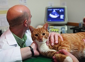
An echocardiogram is performed to evaluate the thickness of the left ventricle, as well as to measure & evaluate the flow of blood and pressures in various heart chambers. Echocardiography is a specialized skill that takes years of practice to properly perform and interpret. Any veterinarian can technically purchase echocardiography equipment, but board-certified veterinary cardiologists have the specialized training, skills, and experience to accurately interpret images and measurements to develop a logical treatment plan for affects cats.
Other non- and minimally-invasive that may be helpful and indicated include:
- Radiography (x-rays) – to evaluate the size of the left atrium, as well as look for evidence of pleural effusion and congestive heart failure
- Electrocardiography (ECG, EKG) – to evaluate the electrical activity of the heart
- DNA testing – blood and cheek swabs from Maine coons and ragdolls can be submitted for analysis
How is HCM treated?
To date there is no treatment that effectively slows progression of hypertrophic cardiomyopathy before the onset of congestive heart failure. Drugs like angiotensin converting enzyme inhibitors (ACEi) and spironolactone have no benefit prior to the onset of CHF. Atenolol, a beta blocker, may reduce the severity of SAM, but does not appear to prolong survival when started prior to onset of CHF.
When CHF is present, patients benefit from diuretic therapy, most commonly a loop diuretic called furosemide (Lasix). Understandable, oxygen supplementation is usually initially indicated. A selective phosphodiesterase inhibitor and calcium sensitizer called pimobendan, as well as ACEi may be beneficial. Cats with meaningful pleural effusion need to have a procedure called a thoracocentesis to evacuate the fluid. Frustratingly there are no medications that effectively breakdown abnormal clot formation. Commonly used drugs – heparin and clopidogrel (Plavix) – can help reduce the likelihood of new clot formation. Furthermore, some cats break down their own clots over time.
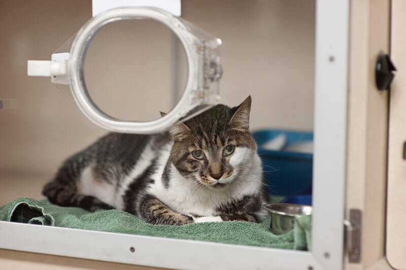
The prognosis for cats with HCM depends on the severity of their disease. Cats with mild or moderate disease may stay at the same level for their entire lives. Cats who develop FATE can suddenly die. Severely enlarged left atria, reduced left atrial function, and severe left ventricular hypertrophy are all negative prognostic factors.
The take-away message about HCM in cats…
Hypertrophic cardiomyopathy or HCM is very common primary heart muscle disease. Many affected cats have no clinical signs, and sudden death is a possible. Therapies are currently aimed at reducing clinical signs associated with sequelae of left ventricular concentric hypertrophy.
To find a board-certified veterinary cardiologist, please visit the American College of Veterinary Internal Medicine.
Wishing you wet-nosed kisses,
CriticalCareDVM

