
The gallbladder plays a critical role in digestion by storing and releasing bile to help break down dietary fats. In dogs, however, this small organ can sometimes develop a life-threatening condition called a gallbladder mucocele. This disease has been increasingly recognized by veterinarians in recent years, and understanding its signs and treatment options can make all the difference for affected pets.
What Is a Gallbladder Mucocele?
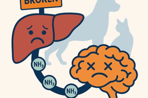
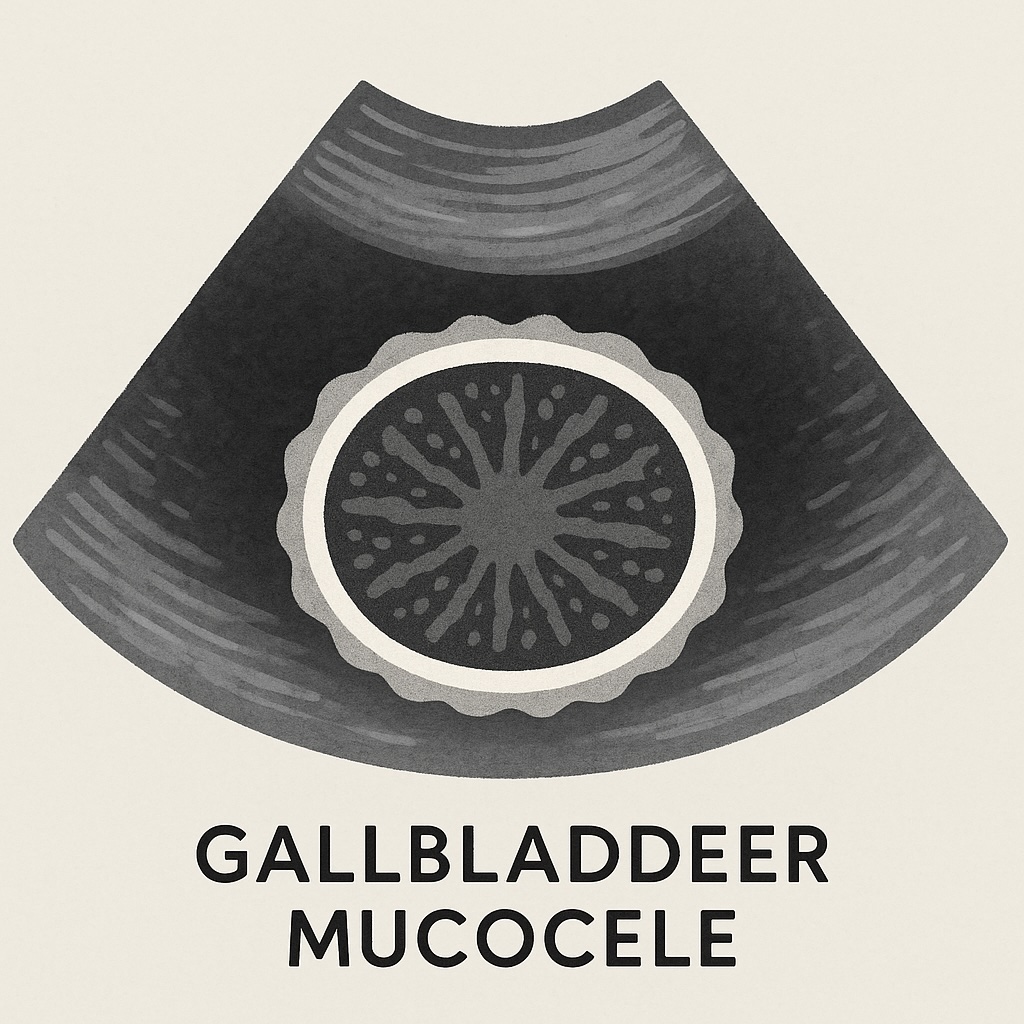
A gallbladder mucocele occurs when the gallbladder fills with an abnormally thick, jelly-like mucus instead of normal bile. This material can block bile flow, stretch the gallbladder, and in severe cases, cause the gallbladder wall to rupture. A rupture can lead to bile leaking into the abdomen, a surgical emergency.
Pathophysiology: How Does It Develop?
While not all of the underlying mechanisms are fully understood, several factors are believed to contribute to mucocele formation:
- Excess mucus production by the gallbladder lining (epithelium), often in response to chronic irritation or hormonal influences.
- Abnormal motility of the gallbladder, preventing it from emptying normally.
- Hormonal and metabolic disease: Dogs with Cushing’s disease (hyperadrenocorticism), hypothyroidism, or hyperlipidemia are at higher risk.
- Genetic predisposition: Certain breeds such as Shetland Sheepdogs, Cocker Spaniels, Border Terriers, and Miniature Schnauzers are overrepresented.
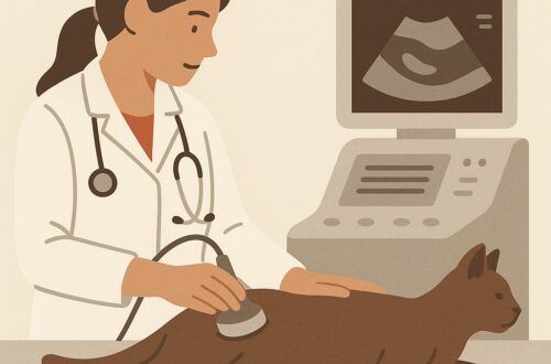
Over time, bile is replaced with this thick mucus, which creates the classic “kiwi fruit” appearance on ultrasound imaging.
Clinical Signs: What Dog Owners May Notice
The signs of gallbladder mucocele can vary, ranging from vague to severe. Common symptoms include:
- Lethargy and weakness
- Decreased appetite or refusal to eat
- Vomiting and abdominal discomfort
- Jaundice (yellowing of the gums, eyes, or skin)
- Increased thirst and urination
- Collapse in severe cases, especially if rupture occurs
Because these signs overlap with many other conditions, diagnosis requires veterinary evaluation.
Diagnostic Testing
Veterinarians use several tools to confirm a mucocele:
- Bloodwork (CBC & chemistry panel)
- May reveal elevated liver enzymes, bilirubin, or cholesterol.
- Evidence of systemic illness (e.g., inflammation, infection).
- Abdominal Ultrasonography
- The current best practice for diagnosis.
- Classic “stellate” or “kiwi fruit” pattern is highly suggestive.
- Helps identify gallbladder wall integrity and signs of rupture.
- Advanced Imaging (CT, MRI) or Bile Culture
- Occasionally used in referral or specialty settings.
Treatment Options
The best practice for treating a gallbladder mucocele in dogs is surgical removal of the gallbladder, known as a cholecystectomy.
- Medical management (ursodeoxycholic acid, antibiotics, hepatoprotectants, dietary modification) has been attempted, but it is rarely rewarding long term. Dogs managed medically face a much higher risk of disease progression, gallbladder rupture, and shortened survival.
- Surgical management provides the best long-term outcome. Dogs can live normal, high-quality lives without their gallbladders once they recover from surgery.
A key consideration is the timing of surgery:
- Elective cholecystectomy (performed when the patient is stable and clinically well) is associated with far fewer perioperative complications and much lower mortality rates.
- Emergency cholecystectomy (performed after rupture or rapid deterioration) carries significantly higher risk, since these patients may already be septic or unstable at the time of surgery.
Whenever possible, veterinarians aim to diagnose early, stabilize patients, and plan surgery electively, giving the patient the best chance of survival.
Prognosis: Why Elective Surgery Matters
When gallbladder mucocele is diagnosed before symptoms develop, and surgery is performed electively, survival outcomes are excellent.
- Elective surgery: Average mortality rate is only about 5%.
- Complications are typically minor, and recovery is smoother.
In contrast, if the dog is already showing clinical signs of disease and requires non-elective (emergency) surgery, the risk of death increases significantly.
- Emergency surgery: Average mortality rate rises to 17–23%.
- About 25% of symptomatic dogs already show gallbladder rupture or bile leakage by the time of surgery. Dogs with rupture have a 2.7-times greater risk of death.
Risk Factors That Worsen Prognosis
Several pre-surgical factors are linked to poorer outcomes in dogs with gallbladder mucocele:
- Advanced age
- Jaundice (hyperbilirubinemia)
- Markedly elevated ALP and ALT levels
- Kidney disease (azotemia)
- Elevated lactate levels (hyperlactatemia)
- Severe or advanced-stage mucocele
- Concurrent endocrine disease such as Cushing’s disease (hyperadrenocorticism)
Why the Immediate Postoperative Period Is Critical
Most deaths occur in the immediate postoperative period, especially in dogs that were unstable before surgery. The most common causes of death include:
- Pulmonary complications such as blood clots (pulmonary thromboembolism), lung injury, aspiration pneumonia, low oxygen (hypoxemia), or respiratory arrest.
- Surgery-related complications like bile peritonitis, biliary obstruction, or sepsis.
- Organ injuries such as acute kidney injury, pancreatitis, peritonitis, or multiple organ dysfunction.
Why Referral to a Specialist Matters
Because dogs with gallbladder mucocele often face significant surgical challenges and require intensive postoperative monitoring, referral to a board-certified surgical specialist and a hospital with access to advanced critical care provides the best chance of survival.
Long-Term Survival
A large study compared outcomes in dogs treated with surgery vs. medical management:
- Surgery group: Median survival ~1,802 days (≈5 years).
- Medical management only: Median survival ~1,340 days (≈3.7 years).
- Medical management first, then delayed surgery: Median survival only ~203 days (≈7 months).
Quality of Life After Surgery
Long-term follow-up shows most dogs thrive after surgery:
- 96% survived to hospital discharge
- 85% were alive at a median follow-up of 1.5 years
- Owners rated post-surgical quality of life as “excellent” (88%) or “good” (12%)
Key Takeaway
Early recognition and elective cholecystectomy offer the best chance for a full recovery. Dogs diagnosed before rupture and stabilized before surgery have dramatically better odds of survival and an excellent long-term quality of life.