I’ve written about spinal cord problems like intervertebral disc disease in previous posts. This week’s post comes as a result of a specific request from one of you, a dedicate follower of CriticalCareDVM.com. Here’s some information about another important spine problem: atlantoaxial instability or AAI. I hope you enjoy reading the information and will share it with other pet owners. Happy reading!

What is AAI?
To understand atlantoaxial instability, you need to have a basic knowledge of spine anatomy. The vertebral column is divided into regions – cervical (neck), thoracic (back), lumbar, sacrum (tail bone), and coccygeal (tail). The first bone of the cervical region – aka C1 – is called the atlas and the second bone – aka C2 – is called the axis. The atlas connects to the skull via the atlanto-occipital joint. The atlas also forms a joint with the axis called the atlantoaxial joint. The latter has a special protrusion called an odontoid process or dens that helps connect the axis to the atlas. This structure allows for greater range of motion between the skull, atlas, and axis.
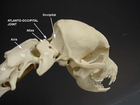
Atlantoaxial instability is characterized by abnormal movement in the neck, specifically between the atlas and axis. The two main causes of AAI are trauma and congenital anomalies. The most common birth defect is an absent or smaller than usual dens (called aplasia and hypoplasia, respectively). Neurologic signs manifest due to compression of the spinal cord caused by the instability.
What does it look like?
Atlantoaxial instability or AAI – also know as atlantoaxial malformation and atlantoaxial subluxation/luxation – is relatively uncommon. There is no sex predilection, and pediatric and young adults are over-represented. This problem is most frequently diagnosed in small breed dogs, including:
- Yorkshire terriers
- Toy & miniature poodles
- Maltese
- Japanese chins
- Chihuahuas
- Pekingnese
- Pomeranians

To date, no cat breed is over-represented. Clinical signs are the result of spinal cord compression, and usually have an acute onset. However, sometimes they are slowly progressive and intermittent, only becoming more obvious after minor trauma like falls. Common clinical signs are:
- Neck pain
- Ataxia (unsteadiness while walking)
- Weakness of all four limbs
- Abnormal postural reactions (e.g.: hopping, wheelbarrowing, paw placement, visual & tactile placing, hemiwalking, extensor postural thrust)
- Paralysis of all four limbs
- Intermittent collapse
- Abnormal carriage of the head
- Breathing paralysis
Watch the video below to view some common clinical signs of AAI in a Yorkshire terrier patient.
Uncommonly, AAI can cause compression of the medulla oblongata in the brain stem, a part of the brain responsible for regulating breathing, heart and blood vessel function, digestion, sneezing, and swallowing. Affected animals may have facial nerve paralysis, vestibular (dizzy) signs, and dysphagia (difficulty swallowing).
How is AAI diagnosed?
Diagnosis of atlantoaxial instability is based on history, clinical signs, breed, age, and diagnostic imaging. Radiography (x-rays) is the most common diagnostic imaging modality used to evaluate patients with suspected AAI. Advanced imaging, including computed tomography (CT scan) and magnetic resonance imaging (MRI), may also be valuable to evaluate resolution of bony abnormalities and identify concurrent soft tissue abnormalities, respectively.
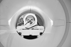
Owners are strongly encouraged to collaborate with a board-certified veterinary neurologist or board-certified veterinary surgeon to help ensure an accurate diagnosis is made.
How is it treated?
Treatment of AAI may be conservative or surgical depending on the severity of a patient’s clinical signs. The former is for pets with mild clinical signs, and involves a minimum of six weeks of strict kennel rest and application of a rigid neck brace. Surgery is recommended for those with meaningful neurologic deficits and for those who don’t respond adequately to conservative therapy. The two types of surgical procedures used to correct AAI are:
- Ventral Fusion – this preferred surgery uses pins, screws, plates, bone cement, and bone grafts to allow physiologic fusion of the atlantoaxial joint.
- Dorsal Fixation – wires, sutures, bands, pins, and bone cement are used to stabilize and align the dorsal spinal processes of the atlas and axis.
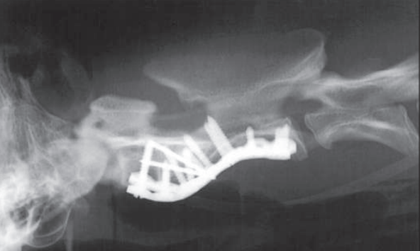
Surgical success rates vary from 60-90%. Regardless of the type of surgery performed, patients require appropriate multimodal pain relief. Once cleared by a veterinary neurologist or surgeon, affected patients often benefit from rehabilitation under the supervision of board-certified veterinary sports medicine and/or rehabilitation specialist or a certified canine rehabilitation practitioner.
The take-away message about AAI in cats & dogs…
Atlantoaxial instability or AAI is a spine problem most commonly identified in pediatric and young adult cats and dogs due to either trauma or congenital defects. Diagnosis is relatively straightforward, and treatment may be conservative or surgical.
To find a board-certified veterinary neurologist, please visit the American College of Veterinary Internal Medicine.
To find a board-certified veterinary surgeon, please visit the American College of Veterinary Surgeons.
To find a board-certified veterinary sports medicine and rehabilitation specialist, please visit the American College of Veterinary Sports Medicine and Rehabilitation.
Wishing you wet-nosed kisses,
CriticalCareDVM


