I find eyes fascinating. They’re beautiful. They have so many unique structures. One of these structures is the nictitating membrane or third eyelid. Occasionally in dogs and cats, the gland associated with this membrane prolapses or slips out of place – a condition commonly known as cherry eye. This week I share some information about this problem. I hope you find it helpful and shareworthy. Happy reading!
Cherry Eye – What is it?
The nictitating membrane or third eyelid is located at the inner or medial aspect of the eye (called the medial canthus). It is made up of conjunctive, a tear-producing gland, and a T-shaped cartilage. The third eyelid has several important functions, particularly:
- Wiping away debris from the surface of the cornea (clear part of the eye)
- Fighting against eye infection
- Producing tears
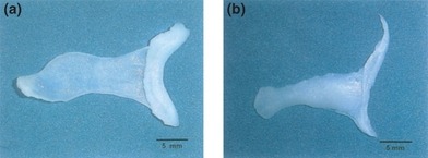
The gland of the third eyelid produces tears. It is located at the base of the nictitating membrane. Under normal circumstances, it is not readily visible.
Cherry Eye – What causes it?
We don’t know what causes of the gland of the third eyelid to prolapse. Prolapse may be unilateral or bilateral. In fact, this problem occurs unilaterally 64% of the time. Bilateral prolapse occurs simultaneously in 41% of dogs. In those with bilateral prolapse that occurs at different times, the second eye prolapses within three months of the first one.
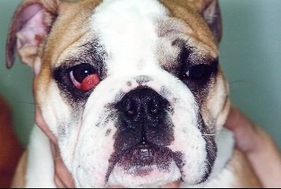
This condition occurs much more frequently in dogs than cats, and does not seem to be painful. There is no sex predilection, and prolapse occurs most commonly in juvenile and young adult patients. Several breeds appear to be predisposed to prolapse, including:
- American cocker spaniel
- Bulldogs (English & French)
- Chinese shar pei
- Boston terrier
- Shih Tzu
- Pekingese
- Neopolitan mastiff
- Newfoundland
- Great Dane
- Beagle
- Bloodhound
- Cane corso
- Lhasa apso
Cherry Eye – How is it diagnosed?
Diagnosis is straightforward. When prolapsed, the gland of the third eyelid appears as a pink or red, rounded mass at the medial canthus. Given the gross appearance, this condition is often referred to as cherry eye because of the resemblance to a cherry pit.

Due to exposure, adjacent conjunctiva is typically very reddened and the affected eye produces mucopurulent discharge. Veterinarians should perform a complete ophthalmic examination, including:
- Schirmer tear test – to assess tear production
- Fluorescein dye test – to check for corneal ulcers
- Tonometry – to check pressure inside the eye
Cherry Eye – How is it treated?
The preferred treatment for prolapsed third eyelid glands is surgical replacement into normal anatomic positioning. There are two general types of surgeries that may be performed to achieve this goal: tacking and pocketing. With the former, the gland is anchored to the adjacent conjunctiva while the latter covers the gland with the surrounding conjunctiva. Tacking procedures are more challenging to perform and require specialized training and expertise. Peri-operative antibiotic and anti-inflammatory eye drops are applied to prevent bacterial infection and reduce inflammation. Pet parents will likely find it helpful to consult with a board-certified veterinary ophthalmologist to determine the best surgical procedure for their fur babies.
With rare exception, the third eyelid gland should not be surgically removed. Surgical removal is often associated with keratoconjunctivitis sicca (KCS or dry eye) and injury to the surrounding cells. Removal is typically only recommended when the prolapse has been chronic, when scar tissue has replaced tear-producing tissue, and KCS is already present. Occasionally veterinarians will recommend surgical removal in older dogs with suspect cancer of the third eyelid gland. Consultation with a board-certified veterinary ophthalmologist is strongly recommended before pursuing surgery to remove the third eyelid gland.
The prognosis for cherry eye is usually good. Understandably, success rates are based on a veterinarian’s experience expertise. Board-certified veterinary ophthalmologists have extensive training in both pocketing and tacking procedures. Factors that have been shown to be negatively associated with outcome are deformation of the T-shaped cartilage, inflammation and enlargement of the gland, and prolonged duration of prolapse.
The take-away message about cherry eye in dogs…
Cherry eye is a term used to describe the non-painful prolapse of the tear-producing gland associated with the nictitating membrane or third eyelid. The prolapsed gland appears as pink or red tissue at the inner point of the affected eye. With prompt recognition and surgical replacement into normal position, affects pets can lead a long, healthy life.
To find a board-certified veterinary ophthalmologist, please visit the American College of Veterinary Ophthalmologists.
Wishing you wet-nosed kisses,
CriticalCareDVM


