Infectious organisms are everywhere. Bacteria, viruses, and fungi threaten our pets every day. Some prefer specific environmental conditions to thrive. Two such organisms are Coccidioides immitis and Coccidioides posadasii, the microbes that cause coccidioidomycosis or Valley fever. This week I hope you’ll enjoy learning more about this important infectious disease in pets. Happy reading!
Coccidioidomycosis – What is it?
Coccidioides spp. are fungal organisms found in the soil in the Lower Sonoran life zone – the southwestern United States (e.g.: southern California, Arizona, New Mexico, Utah, Nevada, and southwest Texas), Mexico, Central America, and parts of South America. Although more prevalent in these regions, pets often travel with their human companions. Thus, technically infected pets may be diagnosed in any geographic location if they’ve previously been exposed to an endemic environment.
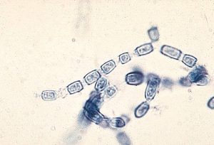
The life cycle of Coccidioides spp. is unique. In soil, a single structure called an anthroconidium germinates to become hyphae that eventually form multiple anthroconidia. Anthroconidia are alternating structures of living and dead cells. They ultimately break into segments at the level of the dead cells and are dispersed in the air or reenter soil to form new hyphae. Pets inhale these aerosolized segments that enter the lungs. In the lungs, they divide to form thousands of endospores that infect lungs and are carried to other parts of the body to cause damage. This process takes 1-3 weeks in dogs, but the fungi can remain in a dormant state for more than three years before clinical signs are noted.
Coccidioidomycosis – What does it look like?
Many dogs living in endemic regions for coccidioidomysis are exposed to the fungal organism, but they don’t develop clinical disease. These dogs may develop a mild cough, but ultimately experience spontaneous remission without treatment. Studies have shown outdoor dogs and those allowed to roam on more than one acre of land were more likely develop disease; similarly, walking dogs on sidewalks in endemic regions conferred a protective effect.
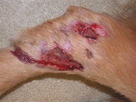
Clinical signs depend on the organ(s) affected by infection, and may include:
- Fever
- Weight loss
- Reduced (or loss of) appetite
- Lethargy
- Weakness
- Lameness
- Enlarged lymph nodes
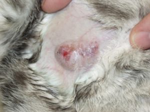
- Draining skin lesions
- Red eyes
- Loss of vision
- Pain
- Seizures
- Abdominal effusion
Coccidioidomycosis – How is it diagnosed?
Veterinarians will ask you many questions about your pet’s medical history. It is essential you tell them if your pet has travelled to an area with endemic coccidioidomycosis. The doctors will also perform complete physical examinations to look for clues about a definitive diagnosis, including enlarged lymph nodes, eyeball changes, abnormal heart rhythms, and lameness.
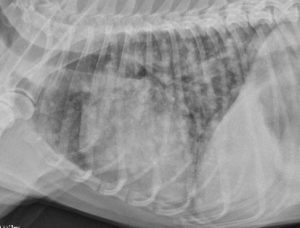
Blood and urine testing should be performed in patients with consistent medical histories and/or physical examinations in an effort to detect the invading fungus and evaluate major organ systems. Additionally, diagnostic imaging in the form of the radiographs (x-rays) are essential. Occasionally, advanced testing like echocardiography (heart sonography), tissue biopsies, cerebrospinal fluid analysis (spinal tap), are recommended. Pet parents may find it helpful to consult with a board-certified veterinary internal medicine specialist to develop a logical and cost-effective diagnostic plan.
Coccidioidomycosis – How is it treated?
With a definitive diagnosis, infected animals need to be treated with anti-fungal medications. Antibiotics have NO effect, as this infection is not caused by bacteria. Drugs that may be prescribed are:
- Ketoconazole | Nizoral®
- Itraconazole | Intrafungol®, Sporonox®
- Fluconazole | Diflucan®
- Amphotericin B (lyophilized)
Prolonged therapy (i.e.: multiple months) antifungal medication is often necessary, and pets should be monitored closely for positive response to treatment. Veterinarians will closely monitor blood and urine to help accurately gauge a pet’s response to therapy and to ensure no adverse reaction to medication.
Pets with meaningful involvement of their lungs and/or other vital internal organs often require around-the-clock hospitalization for critical care, including supplemental oxygen and fluid therapy, until they stabilize. Occasionally, dogs and cats need to have more invasive procedures performed to achieve adequate infection control, including removal of the pericardium (sac around the heart) and/or severely affected eye. Patients with infection localized to the lungs have a good prognosis. Those with disseminated disease have more guarded prognoses with an overall survival rate of 60%. Involvement of the central nervous system and/or bone are worrisome.
The take-away message about coccidioidomycosis in dogs & cats…
Coccidioidmycosis, also called Valley Fever, is caused by the fungal organisms, C. immitis and C. posadasii. Clinical signs are variable, including lameness, lethargy, coughing, and skin lesions. With prompt identification, effective treatment is available.
To find a board-certified veterinary internal medicine specialist, please visit the American College of Veterinary Internal Medicine.
Wishing you wet-nosed kisses,
CriticalCareDVM


