There are many diseases that can affect the lower urinary tracts of dogs and cats – urinary bladder tumors, urinary tract infections, and ectopic ureters. Unfortunately, not all of them can be diagnosed via a simple urinalysis or urine culture. Sometimes we need to look inside the lower urinary tract (LUT). This can be accomplished via a minimally invasive procedure called cystoscopy. This week’s post is dedicated to sharing information about this invaluable diagnostic test, and I hope you find it insightful. Happy reading!

Cystoscopy – What is it?
The fully understand cystoscopy, one should have a basic knowledge of LUT anatomy. The LUT of male dogs is composed of the following components:
- Urethra (tube that connects the urinary bladder to outside world)
- Prostate
- Urinary bladder
In females, the components are:
- Urethra
- Vestibule – the opening into the vagina
- Vagina
- Urethra
- Urinary bladder
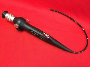
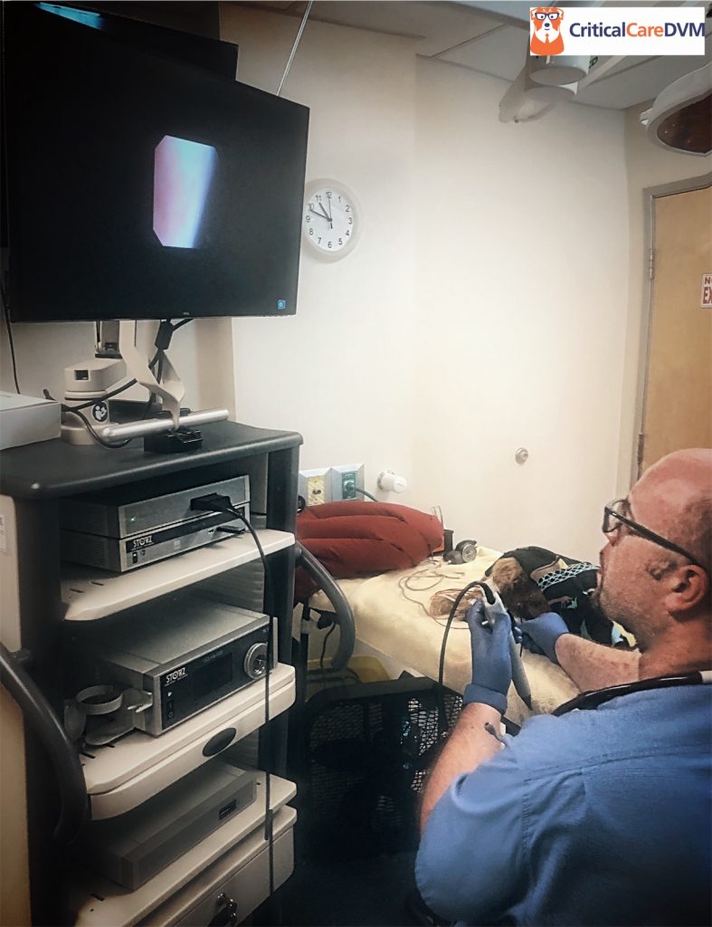
Cystoscopy is a minimally invasive diagnostic imaging modality. Using either a flexible or rigid tube with an attached fiber optic camera and lenses, the instrument is guided to the various parts of the LUT to visualize internal surfaces and obtain tissues.


What should be done before cystoscopy?
Prior to performing this procedure, veterinarians will perform some tests to both screen for possible causes of a pet’s clinical signs and to make sure there are no contraindications for general anesthesia. These tests may include:
- Complete blood count – to evaluate white blood cells, red blood cells, and platelets
- Serum biochemical profile – to evaluate blood kidney values, liver enzymes, electrolytes (i.e.: sodium, potassium), and protein values
- Urinalysis – to screen for evidence of infection, lower urinary tract inflammation and/or bleeding, and inappropriate urine concentration
- Urine culture
- Abdominal imaging (radiographs +/- sonography)
- Urine protein:creatinine ratio (UPC)
- Measurement of urinary bladder residual volume – this volume is the amount of urine left in the urinary bladder after a pet urinates
How is it performed?
Cystoscopy requires general anesthesia to perform. The instrument is lubricated and gently initially inserted in the urethra (males) and vestibule (females). The tube with associated camera/lenses is then advanced through the LUT. Sterile saline is infused into the LUT during the procedure. Filling the urethra and urinary bladder allows optimal visualization.

During the procedure, veterinarians look for anything that appears unusual. The urinary bladder wall should be smooth, and there should not be any blockages in the LUT. During a cystoscopy, veterinarians are able to see:
- Uroliths (stones/calculi in the LUT): Uroliths form from minerals in urine. They usually form when urine does not completely leave the urinary bladder and the minerals in the urine crystallize. If not treated, they can cause pain and lead to hematuria (blood in the urine). Stones can also cause blockages so urine cannot leave the urinary bladder
- Abnormal tissue, polyps, tumors, etc. in any part of the LUT
- Urethral stricture (narrowing of the urethra): Abnormal narrowing may be caused by trauma, infection, tumors, etc.
Below is a video of cystoscopy in a normal female dog performed by a board-certified veterinary internal medicine specialist.
Why is it performed?
Cystoscopy is performed to diagnose, monitor, and/or treat conditions that affect the lower urinary tract, most commonly the urethra and urinary bladder. It’s typically an outpatient procedure and takes less than 15 minutes to complete. Performing cystoscopy requires appropriate training and experience. Board-certified veterinary internal medicine specialists receive extensive instruction in safely, efficiently, and effectively performing this minimally invasive procedure.
During a cystoscopy, veterinarians may be able to treat minor problems such as bleeding in the urinary bladder or blockage in the urethra. Doctors may also use a cystoscopy to:
- Remove small uroliths in the urinary bladder and/or urethra
- Remove or treat abnormal tissue, polyps, and certain tumors
- Inject medication into the urethra wall or the urinary bladder to treat urinary incontinence
Below is a video of cystoscopy in a dog that had a cancerous tumor called a transitional cell carcinoma in the urethra and urinary bladder.
The take-away message about cystoscopy in dogs & cats…
Cystoscopy is a minimally invasive diagnostic imaging procedure used to visualize the various parts of the lower urinary tract. This technique can be used to help accurately diagnose, monitor, and effectively treat a variety of conditions that affect the LUT.
To find a board-certified veterinary internal medicine specialist, please visit the American College of Veterinary Internal Medicine.
Wishing you wet-nosed kisses,
CriticalCareDVM


