An old English proverb states “the eyes are the window of the soul.” I’m quite certain there isn’t anything more soulful than the eyes of a beloved dog or cat. Yet sometimes those beautiful orbs of sight are injured or become diseased. A common eye disorder is glaucoma, and this week I share some information about this painful problem. Happy reading!
Glaucoma – What is it?
Glaucoma is the term used to describe increased pressure within the eye (intraocular pressure or IOP) to a point where the eye can’t function normally. A special structure called the ciliary body produces fluid inside the eye called aqueous humor. This fluid flows through the back of the eye (called the posterior chamber) into the front of eye (called the anterior chamber) via the pupil. Aqueous humor leaves the eye either through an area called the iridocorneal angle (aka drainage angle) or by absorption.
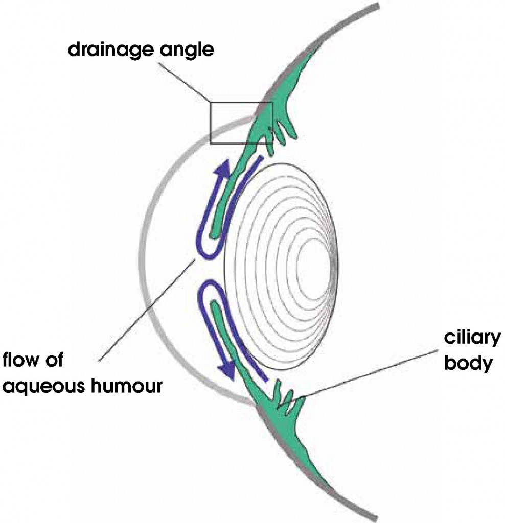
The body is normally able to maintain a normal IOP by delicately balancing the production of aqueous humor with its drainage from the eye. An elevation in IOP occurs when aqueous humor drainage is impaired, creating an imbalance between aqueous humor production and outflow. The impairment of outflow is usually a complication of a pre-existing eye disorder. Elevated IOP causes several major adverse events in the eyes including:
- Disruption of normal nerve signals
- Alteration of blood flow to the eye
- Permanent loss of vision
Glaucoma may be classified into one of two categories: primary glaucoma and secondary glaucoma. The former appears to be breed related, and is often hereditary in nature. One or both eyes may be affected, and is the result of an intrinsic problem of the eye. There is no other ocular disease or disorder. Primary glaucoma may occur at any age, but middle aged dogs (4-9 years of age) are most susceptible. Primary glaucoma is often referred to as either open-angle glaucoma or angle-closure glaucoma depending on the appearance of the iridocorneal angle. Some breeds of dog are over-represented for primary angle-closure glaucoma:
- Cocker spaniels (American & English)
- Malamutes
- Springer spaniels (English & Welsh)
- Basset hounds
- Akitas
- Norwegian elkhounds
- Samoyeds
- Siberian huskies
- Beagles
- Bovier des Flandres
- Chow Chows
- Shar Peis
- Dalmations
- Cairn terriers
- Afghan hounds
Secondary glaucoma is more common than its primary counterpart, and also may affect one or both eyes. It is essentially a complication of another eye disease, such as:
- Anterior lens luxation (displacement of the lens into the anterior chamber of the eye)
- Anterior uveitis (inflammation of the middle layer of the eye)
- Hyphema (blood in the eye)
- Retinal detachment
- Cancer inside the eye
- Iris abnormality
Some cats may develop a unique form of glaucoma called feline aqueous misdirection syndrome (FAMS). With this disorder that typically occurs in older cats, the flow of aqueous humor is misdirected to the back of the eye, resulting reduced drainage to cause IOP to elevate.
Glaucoma – What does it look like?
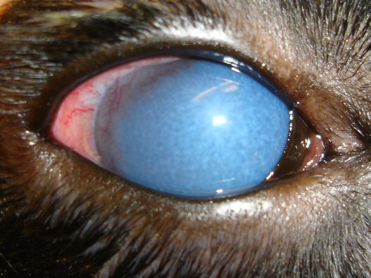
The onset of glaucoma, as well as the rate at which IOP increases, varies significantly and contributes to the severity of the disease and urgency of medical intervention. Changes pet parents may observe at home include:
- Enlargement of the eye
- Tenderness around the eye (or head)
- Red eye
- Cloudy eye
- Discharge from the eyes
- Vomiting
- Diarrhea
- Reduced activity level
Glaucoma – How is it diagnosed?
Measuring IOP in dogs and cats is relatively easy. A veterinarian will use a special instrument to measure IOP. Some primary care veterinarians do not have the instrumentation needed to accurately measure IOP. In such scenarios, families should be referred to a board-certified veterinary ophthalmologist so an affected pet can receive a thorough ophthalmic evaluation.
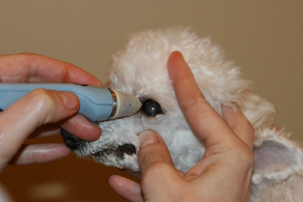
Once elevated IOP has been documented by a primary care doctor, you will likely be referred to a board-certified veterinary ophthalmologist for some additional testing that may include:
- Gonioscopy – a special instrument used to visualize the iridocorneal angle
- High-resolution ultrasonography – a non-invasive modality to evaluate the inner anatomic structures of the eye
- Electroretinography – a minimally invasive test to measure the electrical responses of the retina
- Blood and urine testing to look for underlying causes in patients with secondary glaucoma
Glaucoma – How is it treated?
Patients living with glaucoma may be treated medically or surgically to reduce IOP, preserve vision, and restore patient comfort. Dogs and cats with untreated glaucoma can rapidly go blind. Without timely intervention, vision loss will be permanent. Thus expedient evaluation by a board-certified veterinary ophthalmologist is instrumental in developing the most appropriate treatment plan for an affected patient. Medical management involves the application of eye medications that reduce the amount of aqueous humor in the eye either by:
- Dehydrating the anterior and posterior chambers of the eye
- Reducing aqueous humor production
- Manipulating the iridocorneal angle
- Promoting absorption of aqueous humor
There are various surgical procedures that help reduce IOP and provide improved patient comfort. Potential procedures include:
- Gonioimplantation – a minor procedure to place a filtering device at the level of iridocorneal angle to increase the outflow of aqueous humor from the eye
- Cyclocryoablation – a minimally invasive procedure that freezes all or part of the ciliary body, the part of the eye responsible for making aqueous humor
- Cyclophotocoagulation – a minimally invasive procedure that uses a diode or neodymium:yttrium-aluminum-gamet (Nd:YAG) laser to destroy portions of the ciliary body, the part of the eye responsible for making aqueous humor
When patients fail to respond adequately to medical and/or surgical management and/or when vision loss is permanent, a “salvage” procedure may be recommended simply to ensure an affected pet is as comfortable as possible. These procedures are:
- Enucleation – the surgical removal of the eyeball from a patient with an irreversibly blind and painful eye and/or for those with a cancer inside the eye
- Evisceration with intrascleral prosthesis – a cosmetic alternative to enucleation that involves removing the contents of the eyeball and replacing them with a non-visual prosthetic implant
- Pharmacologic ablation – an injection into the ciliary body to completely iradicate the production of aqueous humor.
Early and aggressive medical and surgical intervention provide patients with the best opportunity for preserving vision. Glaucoma is not curable, and response to long-term medical management eventually diminishes, and patients will typically require subsequent surgical intervention.
The take-away message about glaucoma in dogs and cats…
Glaucoma is defined as elevated pressure within the eye. It is a painful condition that can rapidly result in permanent vision loss if not managed in a timely fashion. Partnering with a board-certified veterinary ophthalmologist is of paramount importance for properly diagnosing the cause of a patient’s glaucoma, as well as maximizing the pet’s comfort and the likelihood of preserving vision.
To find a board-certified veterinary ophthalmologist, please visit the American College of Veterinary Ophthalmologists.
Wishing you wet-nosed kisses,
cgb


