Skin masses are relatively common in dogs. I’ve previously discussed the importance of routine surveillance and early intervention based on the mantras #SeeSomethingDoSomething and #WhyWaitAspirate as advocated by board-certified veterinary cancer specialist, Dr. Susan Ettinger (Dr. Sue Cancer Vet). One group of skin masses is collectively referred to as histiocytic disease, and this week I spend time providing some tangible bits of information about this complex of diseases. Happy reading!
Histiocytic Disease – What are histiocytes?
Histiocytes are special cells of the immune system. There are several types of histiocytes, including:
- Macrophages
- Dendritic cells (i.e.: epithelial/Langerhans, interstitial/dermal, plasmacytoid)
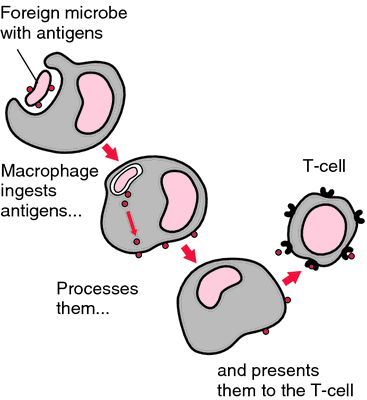
Histiocytes are called antigen-presenting cells or APCs because they come into contact with and process foreign material (called antigens) like bacteria, environmental allergens, and viruses. The most common APCs in dogs are dendritic cells that are found in various layers of the skin. Dendritic cells transport foreign material to the nearest lymph nodes where they present the material to other immune system cells called T lymphocytes. These cells are subsequently stimulated to perform a variety of functions to protect the body.
Histiocytic Disease – Are there different types?
As mentioned earlier, APCs interact uniquely with T lymphocytes. Simplistically speaking, the APCs have keys on their surfaces and the T lymphocytes have the locks on theirs. When the keys line up with the proper locks, all is good. However, when they don’t line up, histiocytic disease can develop.
To date, there are four defined histiocytic diseases in dogs:
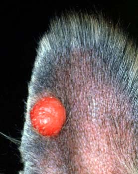
- Cutaneous histiocytoma complex – Lesions are almost always solitary and found in young dogs. They often regress spontaneously, and multiple lesions and metastasis (spread) are exceedingly rare. Langerhans cell histiocytosis is an unusual manifestation of this complex, and is characterized by extensive skin involvement with multiple histiocytomas. Rapid systemic spread and metastasis are possible.
- Cutaneous histiocytosis – This disease is characterized by single or multiple skin masses that tend to wax and wane.
- Systemic histiocytosis – Affected patients have prominent skin lesions, as well as evidence of disease in other organs, including the nose, eyes, spleen, lungs, and bone marrow. The disease is familial in Bernese mountain dogs.
- Histiocytic sarcoma complex – Histiocytic sarcoma and malignant histiocytosis are the components of this highly aggressive cancerous complex. The sarcomas are most commonly found in the spleen, lymph nodes, lung, bone marrow, joint tissue, skin, and the brain. Malignant histiocytosis is very similar, and can be very challenging to differentiate from histiocytic sarcoma. Several breeds are over-represented, particularly Bernese mountain dogs, Rottweilers, Flat-coated retrievers, and Golden retrievers

Histiocytic Disease – How is it diagnosed?
A veterinarian will ask you several questions to get a clear picture of your pet’s medical history. The doctor will also perform a complete physical examination. Skin lesions should be measured and sampled in an attempt to determine their nature. There are a couple methods for sampling skin lumps:
- Fine needle aspiration (FNA) or fenestration: This is the most common technique used to sample cells from a skin lumps/masses. One should note this type of procedure is not a biopsy, but simply an evaluation of cells through use of a microscope – this type of evaluation is called cytology. The skill is very easy to perform and is non-invasive. A sterile small gauge needle (similar to that used to give a vaccine so there is minimal-to-no discomfort for a pet) is introduced into the skin lump/mass with or without an attached syringe. If a syringe is attached to the needle, the plunger of the syringe is pulled back while the needle is in the tissue; this creates suction to pull cells into the needle (aspiration). If no syringe is used, the needle is just gently moved in and out of the skin mass/lump to obtain cells (fenestration). The collected cells are then carefully placed on a microscope slide for evaluation. Below is a video demonstration of fenestration.
- Incisional & excisional biopsies: An incisional biopsy is a surgical procedure in which a small piece of tissue is sampled to identify the composition of a mass. This surgery is usually initially performed if a mass is very large to determine its exact nature before conducting a more invasive surgery to attempt complete removal. An excisional biopsy is a more involved biopsy procedure aimed at removing an entire mass; most small masses are almost always removed in an excisional manner. Your doctor may recommend special testing called immunohistochemical staining be performed on biopsy samples.
Performing these sampling methods is relatively straightforward. Almost all primary care veterinarians are adept at performing FNAs and collecting cytology samples from skin lumps/masses. Many are comfortable procuring both incisional and excisional biopsies too. Occasionally a skin lump/mass will be in a unique location (i.e.: the armpit or groin), and consultation with a board-certified veterinary surgeon may be quite helpful.
Patients with systemic histiocystosis and histiocytic sarcoma complex require an extensive diagnostic investigation that includes:
- Diagnostic imaging (chest & abdominal radiographs/x-rays; abdominal sonography)
- Sampling of the spleen via FNA or fenestration
- Sampling of the bone marrow
Just as for skin biopsy samples, immunohistochemical staining may be performed on spleen and bone marrow samples in an effort to make a definitive diagnosis. Pet parents may find it uniquely helpful to partner with a board-certified veterinary dermatologist and/or cancer specialist to develop a logical and cost-effective diagnostic plan for their fur baby.
Histiocytic Disease – How is it treated?
Treatment of histiocytic disease depends on the type with which a dog has been diagnosed. Patients with a cutaneous histiocytoma may see their mass regress spontaneously. Alternatively, surgical removal is often curative. Patients with reactive and systemic histiocytosis typically require medications that change how the immune system responds. Those living with histiocytic sarcoma complex respond best to chemotherapy, but response is often very short-lived. Collaborating with a board-certified veterinary dermatologist and/or cancer specialist is strongly recommended for patients living with histiocytic disease.
The take-away message about histiocytic disease in dogs…
Histiocytes are cells of the immune system that deliver foreign material to other immune cells in an effort to protect the body. When the interaction between these cells doesn’t go as biologically planned, histiocytic disease can develop. There are several types of histiocytic proliferative diseases in dogs. Clinical manifestation and treatment vary with the type of disease, and thus partnering with a board-certified veterinary dermatologist and/or cancer specialist can be invaluable.
To find a board-certified veterinary cancer specialist, please visit the American College of Veterinary Internal Medicine.
To find a board-certified veterinary dermatologist, please visit the American College of Veterinary Dermatology.
To find a board-certified veterinary surgeon, please visit the American College of Veterinary Surgeons.
Wishing you wet-nosed kisses,
cgb


