Canine athletes, like their human counterparts, are prone to potential musculoskeletal, tendon, and ligament injuries, including cranial cruciate ligament rupture. An under-recognized sporting problem is iliopsoas (il-ē-ō-sō-es) muscle injury. My parents’ dog was recently diagnosed with this condition, so I wanted to dedicate time to explaining it to all of you dog parents out there. I hope you find the information helpful. Happy reading!
Iliopsoas Muscle Injury – What is the iliopsoas muscle?
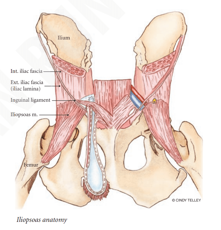
The psoas (sō-es) major muscle arises from multiple thoracic and lumbar vertebrae. It joins with another muscle called the iliacus muscle to become the iliopsoas muscle that ends on the lesser trochanter of the femur (top of the thigh bone). The femoral nerve courses through this important muscle. The major functions of the iliopsoas muscle are:
- Externally rotate the femur (thigh bone)
- Flex the coxofemoral (hip) joint
- Contribute to core stabilization

Iliopsoas Muscle Injury – What does it look like?
There are several types of muscle injuries, and they are classified as one of the following:
- Contusions caused by blunt trauma
- Vascular Compromise
- Lacerations
- Strains
Given its location, the iliopsoas muscle is most commonly injured due to strains. Strains can be acute or chronic in nature, and have variable degrees of severity. In veterinary medicine, muscle strains are categorized as follows:
- Stage I – mild; muscle is inflamed and bruised; often involves delated-onset muscle soreness (DOMS) characterized by the disruption of the Z-band of the sarcomere (a component of a muscle’s contractility mechanism) due to an influx of calcium that propagates an inflammatory response
- Stage II – moderate; muscle is inflamed and some tearing of the muscle sheath has occurred
- Stage III – severe; muscle is inflamed, the muscle sheath has been torn, muscle fibers have been disrupted, and some bleeding into the muscle has occurred
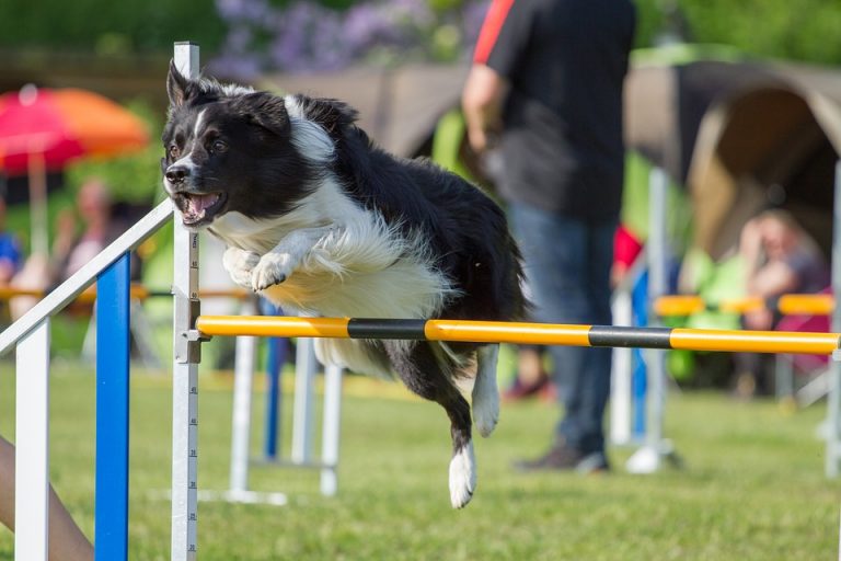
Acute strains are caused by forceful motions (e.g.: turning, twisting) during exercise like jumping or during accidents like slipping. Predisposing factors for acute strains in active dogs are inadequate warm-up, muscle fatigue, and lack of flexibility. Chronic strains are more common than acute ones. Repetitive injuries lead to a cascade of biochemical events in muscle cells that contributes to cramping, formation of myofascial trigger points (“knots” in muscle), and irreversible muscle contracture.
Clinical signs associated with iliopsoas muscle injury are variable. Some dogs show no signs of lameness but may have be hesitant to exercise vigorously and/or have difficulty rising in their pelvic limbs. Others have obvious gait abnormalities due to a reduction in hip extension. There is no age, breed, or sex predilection – any dog may be affected by this problem.
Iliopsoas Muscle Injury – How is it diagnosed?
After obtaining a thorough patient history, a veterinarian will perform a complete physical examination. This should include palpating the iliopsoas muscle to elicit a response from a patient. The ability to fully extend the hip is often compromised. With involvement of the femoral nerve, some dogs may also have a reduced patellar (kneecap) reflex, pelvic limb weakness, atrophy (shrinkage) of thigh muscles, and loss of sensation to the inner thigh. Below is a video showing a dog’s reaction to palpation of its sore iliopsoas muscle.
Diagnostic imaging is appropriate to confirm a clinical diagnosis of iliopsoas muscle injury and to rule out other potential causes of a pet’s clinical signs. This may include radiography (x-rays), muscle ultrasonography, and/or magnetic resonance imaging (MRI). Pet parents will likely find it very helpful to consult with either a board-certified veterinary sports medicine and rehabilitation specialist or surgeon.
Iliopsoas Muscle Injury – How is it treated?
You may be familiar with the acronym RICE – rest, ice, compression, and elevation – for the initial treatment of strains in humans. These same principles technically apply to dogs with iliopsoas muscle injury, and they should be employed for 72 hours after injury to the best of one’s ability. Given the muscle’s location and a dog’s overall anatomy, application of ice and compression, as well as the use of elevation, is limited. Thus, rest is the cornerstone initial treatment.
Injured dogs benefit from multimodal pain medications, non-steroid anti-inflammatory medication, and muscle relaxants. Complimentary therapies, including therapeutic ultrasonography, passive range of motion, stretching and massage exercises, acupuncture, and laser therapy unquestionably help to maximize positive outcomes. Novel therapies, including platelet-rich therapy injections, may also help reduce chronic muscle scarring in some patients. Very rarely is surgery needed for patients with iliopsoas muscle injury. Pet parents are encouraged to partner with either a board-certified veterinary sports medicine and rehabilitation specialist or surgeon to develop the most appropriate treatment plan for their fur babies.
The take-away message about iliopsoas muscle injury in dogs…
Muscle injuries are common in canine athletes. An under-recognized muscular problem in dogs in iliopsoas muscle injury. Easily diagnosed with a complete physical examination and non-invasive diagnostic imaging, affected patients typical positively respond to a combination of interventions, including pain control and rehabilitative exercises.
To find a board-certified veterinary surgeon, please visit the American College of Veterinary Surgeons.
To find a board-certified veterinary sports medicine & rehabilitation specialist, please visit the American College of Veterinary Sports Medicine and Rehabilitation.
Wishing you wet-nosed kisses,
CriticalCareDVM


