Lameness is a common problem in dogs. Muscle injuries like an iliopsoas muscle strain and ligamentous injuries like a cranial cruciate ligament rupture are frequently diagnosed in dogs. Every once and a while, a lameness is more serious than a strain or ligament problem. Sometimes it’s cancer, specifically a cancer in the bone called osteosarcoma. It’s painful. It can be debilitating, especially when recognized too late. So, this week I’m sharing information about this aggressive bone cancer to raise awareness, and I hope you will help me by sharing it with dog parents you know. Happy reading!
Osteosarcoma – What is it?
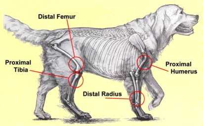
Osteosarcoma is the most common bone tumor is dogs, accounting for 85% of all bone tumors. Bones of the appendicular skeleton – thoracic and pelvic limbs – are most commonly affected. Veterinarians often use the phrase “Away from the elbows & toward the knees” to remember the anatomically predisposed sites, including:
- Distal radius (end of the lower arm bone)
- Proximal humerus (top of the upper arm bone)
- Distal femur (end of the leg bone)

Although commonly involved, bones of the axial skeleton – the core skeletal structures – can be riddled by osteosarcoma. Veterinarians don’t fully understand what causes osteosarcoma. There is evidence this cancer may be attributable to heritable traits. Rapid growth in the first year of life associated with microfractures may predispose to the development of this cancer in later years. Other factors that have been associated with osteosarcoma are radiation therapy, previous fractures (with and without metal implants), and early neutering.
Osteosarcoma – What does it look like?
Large and giant breed dogs are most commonly affected, particularly:
- Rottweilers
- Newfoundlands
- Irish Wolfhounds
- Scottish deerhounds
- Greyhounds
- Great Danes
- Saint Bernards
The most common clinical signs noted in patients with osteosarcoma is lameness. The affected area is also often very painful and firmly swollen. Dogs may chew at the painful regions, and could respond by biting as a defense mechanism if one touches the painful area (or even tries to do so). Lameness may be acute or chronically progressive.
Osteosarcoma – How is it diagnosed?
Veterinarians will initially recommend obtaining radiographs (x-rays) of the tender anatomical location. Osteosarcoma is very aggressive, causing extensive bone destruction (called osteolysis). Occasionally advanced imaging like computed tomography (CT scan) and magnetic resonance imaging (MRI) are needed to detect subtle bone lesions. Ultimately, a bone aspiration or biopsy is required for definitive diagnosis. Biopsies are non-diagnostic only ~10% of patients. It is imperative veterinarians obtain bone samples from the center of the abnormal bone to maximize the accuracy of the results. Since the bone is weakened at the site of sampling, fracturing of the bone is possible – this kind of fracture is called a pathologic fracture because the bone is predisposed to breaking because of disease (not because of a traumatic accident). Reported incident of pathologic fracture post-bone biopsy is less than 1%.

As I mentioned previously, osteosarcoma is a very aggressive cancer. For this reason, complete staging is recommended. Staging is the term used to describe a diagnostic investigation to determine how much the cancer has affected other areas of the body. Staging diagnostic testing may include a complete blood count, serum biochemical profile, urinalysis, chest imaging (radiographs or CT scan), abdominal sonography, and possibly nuclear scintigraphy. A finding of an elevated liver enzyme called alkaline phosphatase (ALP) is a poor prognostic indicator in dogs with appendicular osteosarcoma. Pet parents will likely find it helpful to partner with a board-certified veterinary cancer specialist to develop a logical and cost-effective diagnostic plan.
Osteosarcoma – How it is treated?
Treatment of osteosarcoma centers around treating pain and fighting metastasis (spread of cancer). The most effective way to treat pain associated with appendicular osteosarcoma is removal of the affected area. Unquestionably, osteosarcoma confined to a leg is best treated by amputation of the affected limb. Amputation is a relatively simple surgical procedure, and is well-tolerated by the clear majority of patients. A survey of pet owners whose dogs underwent amputation surgery showed 91% of pet parents perceived no change in their dog’s attitude post-amputation, and almost 75% noted no change in their dog’s activity level. Median survival time for amputation is four months.

Some pet parents are hesitant to have an amputation performed. For these patients, a special surgery called a limb sparing procedure can be performed. During this procedure, the diseased portion of bone is removed, and a bone graft from a donor or a metal spacer is put in its place. The adjacent joint is fused, and chemotherapy is administered post-operatively. A limb sparing procedure should be performed by a board-certified veterinary surgeon. The overall outcome for limb sparing procedures is similar to that for amputation.
Amputation and limb sparing are not viable treatment options for some patients. These dogs require aggressive multimodal pain medications – drugs that target pain relief at different levels to help ensure affected pets are as comfortable as possible. In addition to tradition pain medications, some additional interventions that can provide pain relief are:
- Radiation therapy – this form of therapy can provide good pain control for ~75% of patients for ~2-4 months; when pain recurs, treatment can be repeated
- Stereotactic radiation therapy – a specialized form of radiation therapy that doesn’t cause damage to surrounding tissues; dogs with small bone lesions with minimal destruction appear to have similar outcomes when combined with chemotherapy as pets treated with amputation and chemotherapy
- 153Samarium-EDTMP – a radioactive compound that localizes to bone to cause focal destruction of osteosarcoma cells; approximately 65% of patients experience improvement in pain
- Bisphosphanate infusions – infusion of this class of medication stops the cells that destroy bone (called osteoclasts). When the drug pamidronate was administered monthly, approximately 30% experience meaningful pain relief.
Following surgery, treatment with chemotherapy is recommended because of how rapidly this cancer spreads throughout the body. With platinum based drugs with and without a drug called doxorubicin, median survival is 10-12 months. Dogs with metastases have shorter disease-free intervals (48 days vs 238 days, respectively) and shorter survival times (59 days vs 318 days, respectively) than those without disease spread. Approximately 20% of dogs survive to two years of age.
So far, we’ve been discussing appendicular osteosarcoma. Our discussion wouldn’t be complete without mention of axial osteosarcoma. In general, axial osteosarcoma behaves similarly to the appendicular form. The major difference is a decreased ability to completely remove the boney tumors because of their core anatomical location. Therefore, aggressive local interventions are indicated. Interestingly, osteosarcoma in the maxilla (upper jaw) seems to spread less aggressively.
Osteosarcoma – What’s new?
Veterinary researchers continue to search for newer and more effective therapies for osteosarcoma. Some drugs and targets that may prove effective are:
- Sodium valproate – an inhibitor of histone deacetylase
- EZN3042 – a down-regulator of Survivin, a product of the Birc5 gene
- Auranofin – an inhibitor of thioredoxin reductase
- Hydroxychloroquine – an inhibitor of autophagy (a process of degrading & recycling proteins and other cellular components)
- Co-expression extrapolation (COXEN)
Pet parents are strongly encouraged to partner with a board-certified veterinary cancer specialist to develop the most appropriate treatment protocol for their dog.
The take-away message about osteosarcoma in dogs…
Osteosarcoma is the most common bone tumor that occurs in dogs. Pet parents often bring their canine companions in to see veterinarians because of lameness. Definitive diagnosis requires a bone biopsy, and treatment involves aggressive pain management and interventions to prevent spread of disease.
To find a board-certified veterinary surgeon, please visit the American College of Veterinary Surgeons.
To find a board-certified veterinary cancer specialist, please visit the American College of Veterinary Internal Medicine.
Wishing you wet-nosed kisses,
CriticalCareDVM


Excelente artículo, muy bien redactado y útil a la sociedad
Gracias por leer y compartir con los dueños de los animales.