Lungs are a vital organ essential for delivering oxygen to the lungs and expelling carbon dioxide from the body. In various disease states, the lungs become damaged, and fluid can inappropriately swamp them with fluid. This fluid accumulation in the lungs is called is called pulmonary edema and can dramatically impede oxygenation and ventilation. In this week’s post I share important information about pulmonary edema to help pet owners understand this life-threatening problem. I hope the post helpful and educational. Happy reading!

What is pulmonary edema?
To understand pulmonary edema, one needs to have a basic understanding of lung anatomy. The mouth connects the trachea or windpipe. Inside the chest cavity, there are lung lobes on both the left side and right side. In fact, on the right side, there are four lobes – cranial, middle, caudal, and accessory. There are two lobes on the left – cranial and caudal – with the former subdivided into two subsegments.
The trachea branches into two primary bronchi – one goes to the left side of the chest, the other to right. These bronchi respectively arborize (branch) into smaller and smaller tubules called bronchioles with many of them leading to each lung lobe. Ultimately, the bronchioles terminate into clusters of grape-like sacs called alveoli. A special barrier in the alveoli – called the respiratory membrane – is only three cell layers thick. The layers are:
- Alveolar epithelia (cells that line the alveoli)
- Interstitium (the space between the alveolar & capillary endothelia)
- Capillary endothelia (cells that make up the walls of capillaries)

The respiratory membrane is the site of oxygen and carbon dioxide exchange. The respiratory barrier is damaged by various disease processes via one of two mechanisms: high pressure or increased permeability. Regardless of the mechanism, the result is the same: fluid floods the alveoli to form pulmonary edema to prevent adequate oxygenation and ventilation.
What causes it?
The most common cause of pulmonary edema is heart disease. When heart dysfunction occurs, capillaries develop high pressures inside them. When those pressures become too high, fluid extravasates (leaks out) first into the interstitium and subsequently into the alveoli. Since heart disease is the underlying cause of this type of edema, it’s also called cardiogenic pulmonary edema or congestive heart failure.
Other various disease states can cause capillaries to become more permeable to fluid due to the effect of inflammatory chemicals and enzymes. This type of edema is called permeability edema or non-cardiogenic pulmonary edema, and common diseases associated with it include:
- Acute pancreatitis
- Sepsis
- Acute kidney injury
- Seizures
- Head trauma
- Snake envenomation
- Chest trauma
- Smoke inhalation
- Electrical cord injury

How is pulmonary edema diagnosed?
Cats and dogs with pulmonary edema have difficulty breathing – this difficulty may be mild or severe. Clinical signs associated with mild edema are moist cough, exercise intolerance, lethargy, and/or restlessness. When edema is severe, affected pets cough and may even bring up blood-tinged fluid after retching. They also may hold their elbows away from their chest and stretch their necks. Their mucous membranes (i.e.: gums) often turn a blue/purple/grey color – this is called cyanosis.
Veterinarians will perform a complete physical examination. When they auscultate their heart and lungs (aka use a stethoscope), they often (but not always) hear heart murmurs, abnormal heart rhythms, and/or abnormal lung sounds. If veterinarians are suspicious of pulmonary edema based on pet’s history and physical examination, they will recommend some diagnostic testing that includes:
- Blood work – to screen for major organ dysfunction and electrolyte abnormalities
- Blood pressure measurement
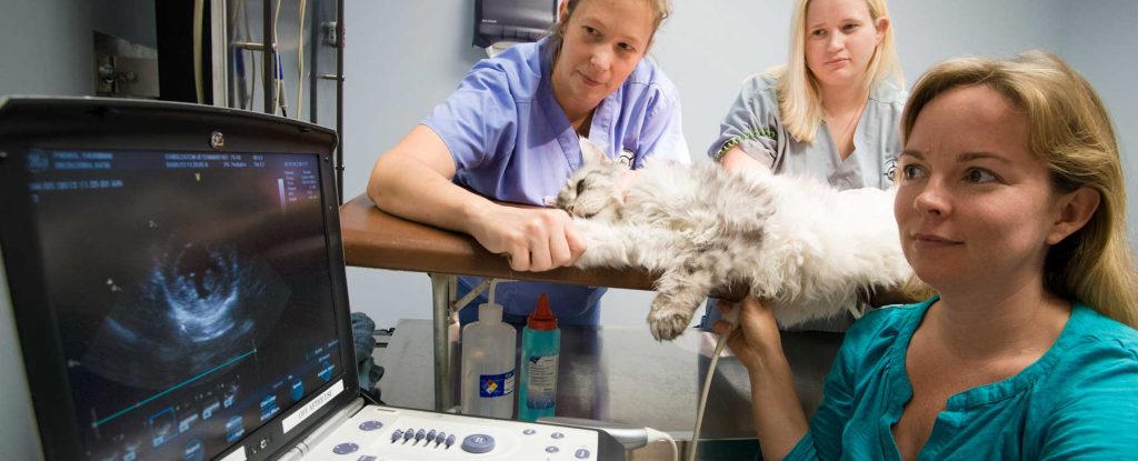
- Diagnostic imaging – chest radiographs (x-rays) and echocardiography (heart ultrasound examination) are essential diagnostic tests for patients with pulmonary edema
- Electrocardiography (ECG/EKG) – to evaluate the electrical activity of the heart
- Pulse oximetry – to assess hemoglobin saturation in the body; hemoglobin is the protein that transports oxygen throughout the body
How is it treated?
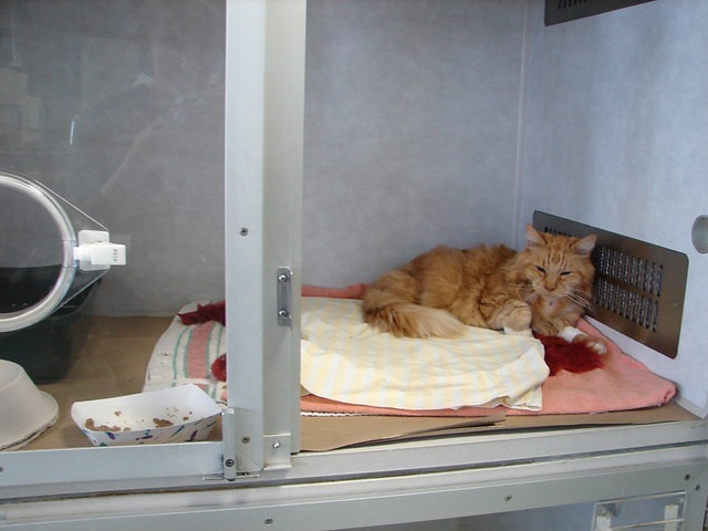
Cats and dogs with pulmonary edema need aggressive medical management. Depending on the severity of their edema, some require temporary hospitalization in a veterinary intensive care unit (ICU) under the direction of board-certified veterinary emergency and critical care specialists. Affected patients need oxygen supplementation and medications to help the body get rid of the inappropriate and excess lung fluid – this class of medication is called diuretics.
For patients with cardiogenic pulmonary edema, other chronic heart supportive medications may be indicated and recommended. Partnering with a board-certified veterinary cardiologist is recommended to develop a chronic treatment plan for patients with cardiogenic pulmonary edema. Patients with non-cardiogenic pulmonary edema may not require chronic interventions if the underlying disease process can be identified and effectively treated/controlled.
The take-away message about pulmonary edema…
Pulmonary edema or water on the lungs can happen with heart disease, as well as other serious medical conditions. Affected patients can develop a life-threatening inability to breathe. Cats and dogs with pulmonary edema need emergency care as soon as possible to maximize the likelihood of a positive outcome.
To find a board-certified veterinary emergency and critical care specialist, please visit the American College of Veterinary Emergency and Critical Care.
To find a board-certified veterinary cardiologist, please visit the American College of Veterinary Internal Medicine.
Wishing you wet-nosed kisses,
CriticalCareDVM


