Eyes are beautiful! The many potential colors of the iris simply make them truly stunning to admire. However, there is one color no one wants to see associated with the eyes – red! In this week’s post, I share some information about red eyes in dogs and cats. Happy ready!
Red Eyes – What causes them?
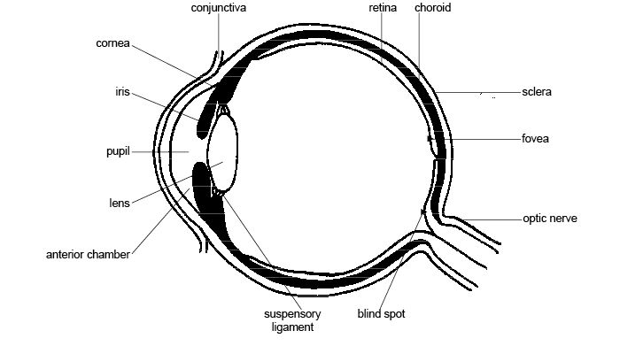
Eyes have several components, and many of them can become reddened when affected by disease. Parts of the eye that frequently become red are:
- Anterior chamber – fluid-filled spaced between the front of the iris and the cornea
- Fundus – the back of the eye that reflects light
- Iris – the colored part of the eye
- Cornea – the clear layer forming the front of the eye
- Conjunctiva – the membrane that covers the outer surfaces of the eyeballs and the inner surfaces of the eyelids
- Sclera/episclera – the white outer layer of the eyeballs
There is a myriad of potential causes for red eyes in dogs and cats. The first step in determining the potential cause(s) is having your family veterinarian perform a complete physical examination on your fur baby. This evaluation includes an extensive examination of the eyes. Particularly helpful findings include:
- Presence or absence of blepharospasm (involuntary closure of the eyelids)
- Presence or absence of discharge from the eyes
- The size of the pupils (holes in the center of the iris)
Patients with blepharospasm look like they are winking at you.
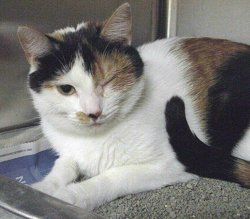
Diseases commonly associated with blepharospasm are:
- Corneal ulcers
- Anterior uveitis (inflammation in the anterior chamber of the eye)
- Acute glaucoma
- Acute lens luxation (dislocation of the lens into either the front or back of the eye)
- Acute keratoconjunctivitis sicca (dry eye)
Certain disorders cause the eyes to produce discharge. The nature of the discharge provides clues as to the cause of a patient’s red eye(s). Similarly, various diseases cause the pupil of the eye to change size. The overall size provides clues as to the potential cause of a patient’s red eye(s). Small pupils (called miotic pupils) are associated with anterior uveitis, while dilated pupils (called mydriatic pupils) are typically the result of chronic glaucoma, retinal and optic nerve disorders, and atrophy (shrinkage) of the iris.
Red Eyes – What diagnostic tests are needed?
Following an ophthalmic physical examination, three non-invasive eye tests are recommended for every patient with a red eye. These tests, frequently called “The Big 3”, are:
- Assessment of tear production
- Measurement of intraocular pressure
- Examination for the presence of a corneal ulcer
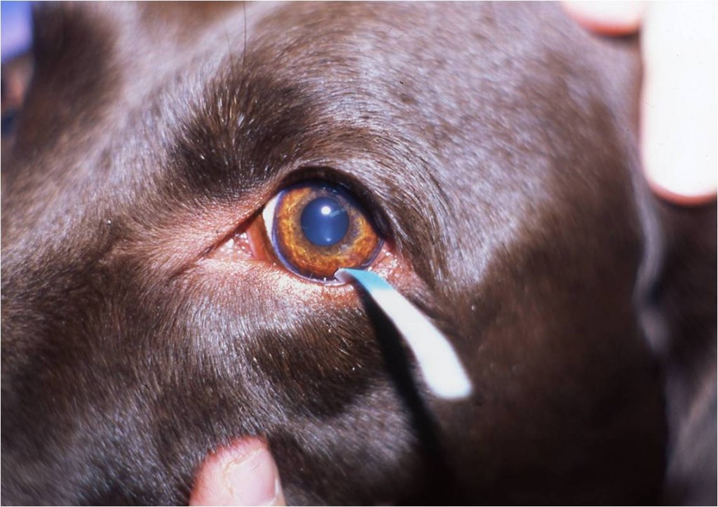
To measure tear production, a veterinarian performs a test called a Schirmer Tear Test (STT). A small test strip is placed within a part of the eye called lower conjunctival fornix. The test strip is left in place for exactly one minute. Tear production is visualized by dye migration down the strip.
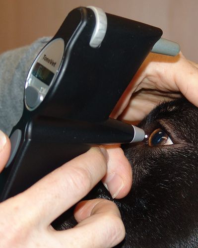
Intraocular pressure or IOP is the fluid pressure inside the eye. Some diseases, like glaucoma, are characterized by elevated intraocular pressure. Others, including anterior uveitis, are associated with decreased IOP. To measure intraocular pressure, veterinarians use a technique called tonometry. There are different types of tonometry, and the three most commonly used in veterinary medicine are:
- Rebound tonometry – uses a magnetized probe that bounces off the cornea when the cornea is touched with the probe
- Applanation tonometry – measures the counter pressure when the probe touches the cornea
- Impression/Indentation tonometry – measure the depth of a corneal indentation made by a small plunger carrying a known weight; the higher the IOP, the harder it is to push against and indent the cornea. The movement of the plunger is measured using a calibrated scale.
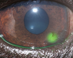
A veterinarian can check for defects in the corneas by applying a special dye called fluorescein. When a lesion like an ulcer is present, the dye is visible with a special light.
Depending on the results of “The Big 3” some additional testing may be recommended. Blood and urine tests may be performed to screen for systemic disorders. A veterinarian may measure a patient’s blood pressure to look for hypertension (high blood pressure). Some other potential eye tests include:
- Ultrasonography
- Rose bengal staining – a special stain to evaluate defects in the cornea
- Slit lamp examination – a instrument that can focus a special light source into a thin sheet of light
- Gonioscopy – a non-invasive examination of the area where fluid drains out of the eye
The eyes are unique organs, and a thorough examination is essential to identify the underlying cause(s) of red eyes. As you have likely figured out, some special instrumentation is needed to perform a proper ophthalmic evaluation. Thus partnering with a board-certified veterinary ophthalmologist is uniquely beneficial for maximizing the preservation of an affected pet’s vision.
Red Eyes – How are they treated?
Effective treatment of red eyes requires an accurate diagnosis. The medication(s) required for a patient living with glaucoma are vastly different than those for a patient with a corneal ulcer. Your family veterinarian will likely recommend referral to a board-certified veterinary ophthalmologist to ensure your pet receives the most appropriate care.
The take-away message about red eyes in dogs and cats…
Pet parents should immediately seek veterinary medical attention should they observe red eyes in the fur baby. There is a plethora of disorders that can cause the reddening of eyes. Efficient diagnosis is crucial. Working with a board-certified veterinary ophthalmologist can help ensure an affected patient is both diagnosed accurately and treated appropriately.
To find a board-certified veterinary ophthalmologist, please visit the American College of Veterinary Ophthalmologists.
Wishing you wet-nosed kisses,
Cgb


