Respiratory distress in dogs and cats is stressful for all involved – pet, owner, and even veterinarians. In previous posts I’ve shared information about various breathing problems, including kennel cough and aspiration pneumonia. This week I wanted to spend some time discussing a truly life-threatening respiratory problem – spontaneous pneumothorax. So, I hope you enjoy the read and find the content informative. Happy reading!

Spontaneous Pneumothorax – What is it?
To best understand what a spontaneous pneumothorax is, one first needs to have a basic understand of the anatomy of the chest cavity. The border of the chest cavity is the rib cage. Inside the chest are important structures like the heart and the various lung lobes. Under normal circumstances, the lungs lie directly adjacent to the rib cage. The potential space between them is called the pleural space. Under normal circumstances there is no space between the chest wall and the lungs because of the physiologic negative pressure in the chest cavity. For various reasons, when negative pressure is lost, air and/or fluid can accumulate in the pleural space – to learn more about pleural space disease in general, click here.
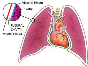
A spontaneous pneumothorax is a condition in which air accumulates in the pleural space without a history of trauma or penetration of the thoracic cavity. In the absence of trauma or chest wall penetration, air enters the pleural space via one of two routes:
- Airway – compromise of the trachea (windpipe) and/or lung tissue can cause air to enter the pleural space
- Esophagus – compromise of this tubular structure that connects the mouth to the stomach can cause air to leak into the pleural space, as well as under the skin and into a space in the middle of the chest cavity called the mediastinum
Major potential causes of problems in the airway and esophagus include:
- Bullae & blebs (airway only) – essentially air blisters that abnormally form in lung lobes
- Cancer
- Infections (bacterial, fungal)
- Heartworm disease
- Pulmonary thromboembolism
Spontaneous Pneumothorax – What does it look like?
There is age or sex predilection for spontaneous pneumothorax. Any breed of dog may be affected, but Siberian huskies are over-represented. Common clinical signs include:
- Increased respiratory rate characterized by short & shallow breaths
- Difficulty breathing
- Anxiety
- Blue / grey / purple gums (called cyanosis)
- Intermittent coughing
- Neck extension
- Elbow abduction (pointed away from the rib cage)

Spontaneous Pneumothorax – How is diagnosed?
When a patient is presented in respiratory distress, the veterinarian will perform a primary survey; that is, they will ensure the airway is patent, use a stethoscope to listen to the heart and lungs, and assess pulse rate and quality. When a pneumothorax is present, one can’t hear lung and heart sounds well.
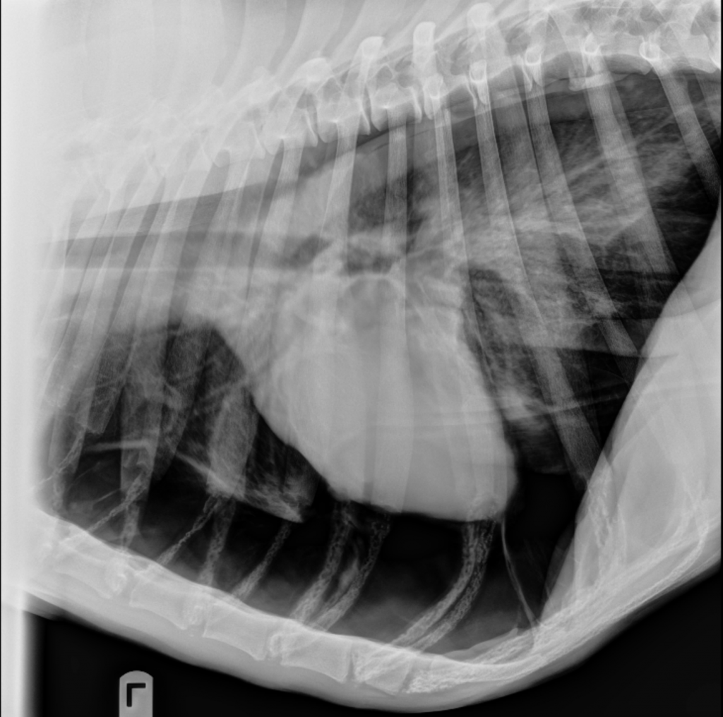
Confirmation of pneumothorax requires diagnostic imaging, most often in the form of radiographs (x-rays). It is important to note radiographs typically only identify the presence of pleural space disease. They rarely identify the underlying cause, and further imaging like computed tomography (CT scan) is needed. Pet parents may find it helpful to partner with a board-certified veterinary surgeon to develop the most logical and cost-effective diagnostic plan.
Spontaneous Pneumothorax – How is it treated?
Patients with spontaneous pneumothorax in respiratory distress initially require a minimally invasive procedure called a thoracocentesis to evacuate air from the pleural space (see video below).
During the procedure, a small needle or catheter is inserted through the chest wall into the pleural space to remove the abnormally accumulated air. Removal allows the lungs to expand more normally, easing the work of breathing for the affected pet. If air re-accumulates rapidly and/or if multiple thoracocenteses are required, a veterinarian will recommend placing a special temporary tube called a thoracostomy tube (aka chest tube) into the pleural space to allow for intermittent or continuous suction of air.
Patients with spontaneous pneumothorax should receive supplemental oxygen therapy until their breathing challenges have been resolved. Some patients can be successfully treated without surgery. However, most patients with spontaneous pneumothorax require surgical intervention. There are three different surgical approaches to the chest cavity (called thoracotomies), and pet parents should consult with a board-certified veterinary surgeon to determine the most appropriate surgery for their pet. The prognosis ultimately depends on the underlying cause of spontaneous pneumothorax. With surgery, the recurrence rate is ~3% and up to 50% without surgery. The mortality is 12% and >50% with surgery and medical management, respectively.
The take-away message about spontaneous pneumothorax…
Spontaneous pneumothorax is potential cause of life-threatening respiratory distress in dogs and cats. Efficient identification, acute stabilizing care, and surgical management are typically needed to maximize the likelihood of a positive outcome.
To find a board-certified veterinary emergency and critical care specialist, please visit the American College of Veterinary Emergency and Critical Care.
To find a board-certified veterinary surgeon, please visit the American College of Veterinary Surgeons.
Wishing you wet-nosed kisses,
CriticalCareDVM


