I’ve previously written about one of the most common problems for which pet owners bring their pets to be evaluated by a veterinarian – otitis externa (aka an ear infection). Unfortunately, some of our furry friends don’t adequately respond to medical interventions. This small population of patients needs surgery. In this week’s post I share information about the recommended surgical intervention for these cats and dogs. Happy reading!

Otitis Externa Refresher
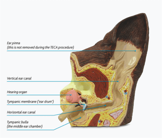
A cat’s/dog’s ear has three major sections: the outer ear, middle ear, and inner ear: The inner ear connects to the brain and contains nerves that help with hearing and balance. Causes are generally divided into predisposing factors (e.g.: swimming, humidity, ear canal trauma), primary factors (e.g.: ear mites, food allergies, polyps, etc.), and perpetuating factors (e.g.: bacteria, fungi). Treatment is directed at addressing both primary and perpetuating factors. Understandably, the therapeutic approach can be quite variable given the myriad of factors that influence this disease. For some dogs, recurrent and chronic inflammation and infection damages the outer ear too badly. Resolution of outer ear disease even with proper cleaning and systemic and topical medications is simply not possible. Such patients need surgery.

Which pets need surgery?
Generally speaking, surgical intervention for otitis externa should only be considered for those patients who have failed to adequately respond to non-surgical management. For pets with end-stage otitis externa, the surgical procedure of choice is a total ear canal ablation and bulla osteotomy (TECA-BO). Specific ear changes for which a veterinarian may recommend a TECA-BO are:
- Calcification of the cartilages within the external ear canal
- Granulomatous proliferations within the external ear canal
- Abscess formation in the tissue surrounding the affected ear
- Rupture of tympanic membrane (ear drum)
- Evidence of middle ear infection (based on computed tomography (CT scan)
- Cancerous growths within the external ear canal
Prior to surgery, a thorough patient assessment is vital. Veterinarians will review a patient’s complete medical records and subsequently perform a thorough physical examination. Pre-anesthetic blood and urine tests (i.e.: complete blood count, biochemical profile, urinalysis, coagulation profile) should be performed just as they would be for human patients undergoing anesthesia and surgery. Advanced diagnostic imaging – typically computed tomography / CT scan – is uniquely beneficial to both confirm end-stage external ear canal disease and assist with surgical planning.
What is a TECA-BO?
Total ear canal ablation with bulla osteotomy is the surgical removal of external ear canal and middle ear. The pinna (ear flap) remains intact. Under general anesthesia, a veterinarian makes an incision and dissects / cuts through the underlying tissue and cartilage so the external ear canal can be removed as one intact cylinder. The tympanic membrane (ear drum) and bones of the middle ear are also removed.
The middle ear – also known as the tympanic bulla – is thoroughly flushed of any infected material. The bone in this region is also curetted or scrapes to ensure all infected material is removed. After thoroughly flushing this area, a sample is collected from this site for culture. A temporary surgical drain is placed to facilitate drainage of any remaining fluid.
Please watch the video below to see a TECA-BO performed on a dog (warning: graphic images).
This surgical procedure is typically performed by a board-certified veterinary surgeon; however, some primary care veterinarians may be comfortable performing this surgery.
Potential Complications
There are inherent risks of general anesthesia and any surgery. These include blood pressure changes, bleeding, incision site complications, abnormal heart rhythms, and even catastrophic adverse events, including death. Thankfully, the risk of these issues is low.
Risks specific to the TECA-BO procedure include:
- Damage to the blood supply of pinna (ear flap) – although uncommon, the margins of the ear flap may die necessitating a follow-up surgery to remove the dead tissue
- Damage to a nerve bundle called the sympathetic trunk, resulting Horner’s syndrome.
- Damage to the facial nerve, leading to facial nerve paralysis – if this occurs, it is usually temporary and typically resolves without treatment; in rare cares facial nerve paralysis can be permanent.
- Chronic incision drainage due to residual infection – this is only reported in 3-15% of patients and usually resolves with antibiotic treatment. Rarely, a second surgery is needed.
Pet owners often ask about hearing loss following TECA-BO. Understandably, hearing impairment is possible after this surgery. However, many cats and dogs with end-stage otitis externa have pre-existing hearing deficits so appreciable change post-operatively is not noticeable.
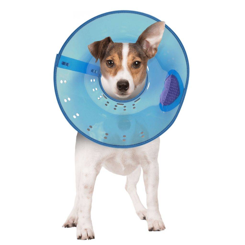
Post-operatively, patients will need to wear an Elizabethan collar, otherwise known as the cone of shame. This is essential to prevent them from traumatizing their healing incision. Patients will be discharged with appropriate pain medications and antibiotic therapy. The overall prognosis is very good, as pets experience relief from chronic pain and families no longer have to manage their pet’s chronic ear infections.

The take-away message about surgery for otitis externa…
Surgery is required for pets with otitis externa who don’t adequately respond to medical therapies. Total ear canal ablation and bulla osteotomy can provide permanent pain relief affected cats and dogs.
To find a board-certified veterinary surgeon, please visit the American College of Veterinary Surgeons.
Wishing you wet-nosed kisses,
CriticalCareDVM


