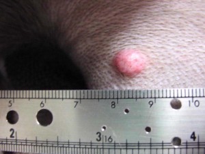
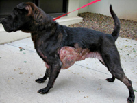
A common reason for a pet parent to bring his/her pet to a family veterinarian is because s/he has discovered a new skin lump/mass on his/her dog or cat. This lump can be big or small. It may cause the pet discomfort or may truly be an incidental finding. Nevertheless determining if a mass is cancerous or not is essential. And you may ask, “Which lumps should be evaluated by my family veterinarian?” A group of board-certified veterinary oncologists (cancer specialists) recommends pets be evaluated by their family veterinarian when a lump/mass has been present for more than a month and/or if it is bigger than a pea. Early identification of skin cancers can be life-saving!
(image courtesy of Bowen Road Animal Clinic; www.arlingtonvet.com)
Sampling a skin lump/mass…
There are a few methods of sampling skin lumps to gain a definitive diagnosis, including:
Fine needle aspiration (FNA) or fenestration: This is the most common technique used to sample cells from a skin lumps/masses. One should note this type of procedure is not a biopsy, but simply an evaluation of cells through use of a microscope – this type of evaluation is called cytology. The skill is very easy to perform and is non-invasive. A sterile small gauge needle (similar to that used to give a vaccine so there is minimal-to-no discomfort for a pet) is introduced into the skin lump/mass with or without an attached syringe. If a syringe is attached to the needle, the plunger of the syringe is pulled back while the needle is in the tissue; this creates suction to pull cells into the needle (aspiration). If no syringe is used, the needle is just gently moved in and out of the skin mass/lump to obtain cells (fenestration). The collected cells are then carefully placed on a microscope slide for evaluation.
Here is a link to a video demonstrated fenestration of one of a dog’s lymph nodes: http://youtu.be/0eH5NRzK7QE
NOTE: Even though the video is entitled ‘Fine Needle Aspirate’ and the doctor speaking uses the term aspiration, the procedure depicted is called fenestration because no syringe was attached to the needle while the needle was in the tissue.
Incisional & excisional biopsies: An incisional biopsy is a surgical procedure in which a small piece of tissue is sampled to identify the composition of a mass. This surgery is usually initially performed if a mass is very large to determine its exact nature before conducting a more invasive surgery to attempt complete removal. An excisional biopsy is a more involved biopsy procedure aimed at removing an entire mass; most small masses are almost always removed in an excisional manner.
Performing these sampling methods is relatively straightforward. Almost all primary care veterinarians are adept at performing FNAs and collecting cytology samples from skin lumps/masses. Many are comfortable procuring both incisional and excisional biopsies too. Occasionally a skin lump/mass will be in a unique location (i.e.: the armpit or groin), and consultation with a board-certified veterinary surgeon may be quite helpful.
(image courtesy of Tracy Geiger, DVM, DACVIM)
What happens to the sample?
If your pet’s primary care doctor performs a FNA for cytology, s/he will likely look at the sample his/herself in the hospital. Indeed I encourage this to promote continued learning, and this initial evaluation can be potentially helpful, providing preliminary information. With that being said, I strongly believe a board-certified veterinary clinical pathologist should always evaluate all cytology samples. I look at cytology samples multiple times a day. Why? I find cytology fascinating. Quite frankly cells are beautiful. But I’m not a board-certified veterinary clinical pathologist. I’m not an expert in microscopic cell evaluation, and neither is your family veterinarian. If you had a skin lump/mass sampled for cytology, would you want your primary care physician evaluating the cells to determine if cancer was present or would you prefer an expert in microscopic cell evaluation looks at them? I suspect most of you reading this would prefer the latter, and I would argue the same care should be provided to every pet.
Biopsy samples should always be submitted for evaluation by a board-certified veterinary pathologist – this assessment is called histopathology. When I was attending veterinary school at Cornell University, every single pathologist drilled a mantra into my head:
If a mass is worth taking off, it is worth finding out what it is!
What does this mean? It means if I’m going to ask a patient to undergo general anesthesia and surgery to remove a skin lump/mass, I want to know what it is. Tissue samples are always submitted to a board-certified pathologist, an expert in evaluating biopsy samples. This probably sounds like common sense (or at least I hope it does), but for various reasons sample submission doesn’t always happen. Sometimes parents don’t authorize histopathology. Some primary care veterinarians make an assumption that a lump/mass is benign and choose not to submit a biopsy for histopathology. This has always perplexed me because the only way to determine if a biopsy is benign or malignant is to perform histopathology, period!
The take-away message about skin lumps…
Skin masses larger than a pea and/or those that have been present for more than one month should always be sampled. Sampling may be performed via fine needle aspiration/fenestration or via surgery to obtain an incisional or excisional biopsy. Your pet’s family veterinarian will help you determine the most appropriate technique for your fur baby. A consultation with a board-certified veterinary surgeon may be helpful for large skin lumps/masses and/or for those located in unique locations. Cell and tissue samples should always be evaluated by a board-certified veterinary clinical pathologist or pathologist, respectively. Please visit www.seesomethingdosomething.net and https://www.facebook.com/seesomethingdosomethingcancer to learn more about diagnosing skin lumps/masses in dogs and cats.
To find a board-certified veterinary surgeon, please visit the American College of Veterinary Surgeons.
To find a board-certified veterinary oncologist, please visit the American College of Veterinary Internal Medicine.
To learn more about board-certified veterinary clinical pathologists and pathologists, please click here.
Wishing you wet-nosed kisses,
cgb


