Have you been told your dog or cat needs biopsies from the gastrointestinal tract? Does this scare you because you’re concerned about your fur baby’s comfort level, anesthesia and recovery? Have no fear because in this week’s post you will find answers to your questions about gastrointestinal biopsies! I am excited to turn Because Pets Can Have Specialists Too over to guest contributor Dr. Fiona Park, another board-certified veterinary internal medicine specialist. She thoroughly discusses the benefits and limitations of two techniques for obtaining biopsies from the gastrointestinal tract of dogs and cats. Happy reading!
Gastrointestinal biopsies – when are they needed?
One of the tests sometimes used to help investigate problems such as chronic vomiting and chronic diarrhea in dogs and cats is biopsy of the gastrointestinal tract. This will often be recommended after simpler tests (such as blood/urine tests, fecal/stool analyses, radiographs/x-rays, ultrasonography) have eliminated other causes of these clinical signs.
Gastrointestinal biopsies – how are they collected?
When biopsies are obtained from the gastrointestinal tract, samples are collected from the stomach, small intestine and/or large intestine. These tissues are subsequently submitted to a laboratory for analysis by a board-certified veterinary pathologist (this is called histopathology). There are two ways a veterinarian can obtain biopsies from the gastrointestinal tract:
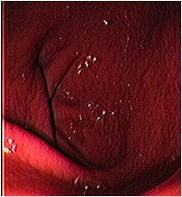
- Endoscopic biopsies – performed using an endoscope, a type of flexible tube attached to a video camera
- Surgery – performed by making an incision into the abdomen (laparotomy) or sometimes “keyhole” abdominal surgery (laparoscopy)
Both of these methods have their own advantages and disadvantages.
Endoscopic biopsies:
Advantages of endoscopic biopsies:
- Endoscopy is a relatively non-invasive procedure: although patients require general anesthesia, recovery from the procedure is rapid for most patients. As the endoscope is introduced to the body through the mouth and/or anus/rectum, there is no incision to heal.
- The inside lining (mucosa) of the esophagus, stomach, parts of the small intestine and large intestine (colon) can be visually inspected at the time of the endoscopy.

- Endoscopic biopsies can safely be obtained from the colon (see below).
- Endoscopic biopsy sites heal very quickly.
- Endoscopic biopsies can be obtain in cats and dogs with low blood protein levels without increased risk of complications.

Disadvantages of endoscopic biopsies:
- Only a limited portion of a dog or cat’s small intestine can be accessed using an endoscope. Most endoscopes are 1-1.5 meters (~40-60 inches) long while the small intestinal tract of dogs and cats is much longer. Thus this may be an issue if the disease only affects a certain area of the small intestine that is out of reach of the endoscope.
- Endoscopic biopsies come from only the innermost layer of the wall of the stomach and intestine (called the mucosal layer). This may be an issue if the disease has only caused changes in the deeper layers of these organs (called the submucosa, muscularis, and serosa).
- The quality of endoscopic biopsies can vary and is highly dependent on the expertise of the veterinarian performing the procedure. For this reason, it may be beneficial to work with board-certified veterinary internal medicine specialists who have undergone extensive and specialized training in endoscopic procedures.
Surgical (full thickness) biopsies:
Advantages of full thickness (surgical) biopsies:
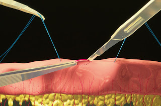
- A sample that includes all the layers in the stomach or small intestine can be obtained. This means diseases that only affect the deeper layers of the stomach or small intestine can still be readily diagnosed.

 TOP: Illustration of a full thickness biopsy of the intestinal tract BOTTOM: Illustration showing all four layers of the intestinal are biopsied with surgical biopsies. Photo courtesy of board-certified veterinary surgeon Dr. Stephen J. Birchard
- During surgery all three segments of the small intestine (called the duodenum, jejunum, and ileum) can be evaluated and biopsied. Furthermore any areas of bowel that are determined to be abnormal from the outermost layer (for example thicker than normal) can be specifically sampled.
- Biopsies can also be obtained from lymph nodes and other organs in the abdomen, most commonly the pancreas and liver, at the time of surgery. This is not possible during endoscopy, and may be important in some cats and dogs with cancer of the gastrointestinal tract.
Disadvantages of full thickness (surgical) biopsies:
- Although both methods of obtaining gastrointestinal biopsies require general anesthesia, recovery time is often longer following surgery than following endoscopy. Accordingly pets undergoing surgery are hospitalized longer compared to those who have an endoscopic procedure performed. Furthermore your pet’s exercise must be restricted until the surgery site has healed completely. If laparoscopy is possible, then recovery time may be reduced.
- Some dogs and cats with gastrointestinal problems have decreased blood protein levels. These patients have a higher risk of complications after full thickness biopsies, as the biopsy sites may not heal as efficiently as they should. These complications can be life-threatening and/or require a second surgery to remedy.
- Full thickness biopsies of the large intestine (colon) are rarely performed because there is a high risk of complications.
The take-away message about gastrointestinal biopsies…
The decision to obtain gastrointestinal biopsies via endoscopy or surgery is an important one. Many factors influence influence a veterinarian’s recommendation for biopsy modality. For this reason, it is crucial you spend an adequate amount of time discussing this decision with your family veterinarian. Additionally you may find it uniquely beneficial to consult with a board-certified veterinary internal medicine specialist, especially if you would like to pursue endoscopy.
I hope you enjoyed Dr. Park’s fantastic post. I love providing you with opinions from other board-certified veterinary specialists, and I plan to welcome more guest bloggers from the world of specialty veterinary medicine. I sure hope you like the variety! Remember pet parents deserve to know this information, and I hope you will consider sharing it with others you know. Of course you can more useful information at my social media sites:
- Twitter: www.twitter.com/CriticalCareDVM
- Facebook: www.facebook.com/CriticalCareDVM
- Tumblr: www.CriticalCareDVM.tumblr.com
- Instagram: www.instagram.com/CriticalCareDVM
- Pinterest: https://www.pinterest.com/CriticalCareDVM
To find a board-certified veterinary internal medicine specialist, please visit the American College of Veterinary Internal Medicine.
To find a board-certified veterinary surgeon, please visit the American College of Veterinary Surgeons.
Wishing you wet-nosed kisses,
cgb
Meet Dr. Fiona Park
Dr Fiona Park BVSc (dist), DVSc, DACVIM (Small Animal Internal Medicine) is a both an Australian small animal internal medicine specialist and a Diplomate of the American College of Veterinary Internal Medicine. Fiona started her specialist training in internal medicine initially as an intern and subsequently as a resident at the University of Guelph Ontario Canada. On achieving specialist status through the American College of Veterinary Internal Medicine, Fiona returned to Australia, obtained her Australian specialist status and started working at the Veterinary Referral Hospital in Sydney. Fiona has a range of publications with a specific interest in disorders of blood coagulation and endocrinologies. Fiona is active in veterinary post graduate training through provision of continuing education lectures and welcomes veterinarians to the hospital to participate in both case rounds and journal reviews.
In addition to accepting referral internal medicine patients Fiona assists general practitioners in the management of their own cases by providing valuable support and advice regarding the interpretation of pathology results. For Fiona having had the privilege of specialist training (and the associated hard work!) it represents an opportunity to give back to people and their pets the opportunity of a healthy and happy life together.


