
Each of us knows that wonderful feeling of looking into the eyes of our fur babies, sensing the non-verbal love we cherish from them. But what do you do when your pet’s eyes appear red? I’m not talking about metaphorical red but rather true physical red that indicates a medical problem is present or brewing. This week I discuss the common causes of “red eye” in dogs and cats, as well as the tests that should be performed on affected animals.
Red eye – potential causes
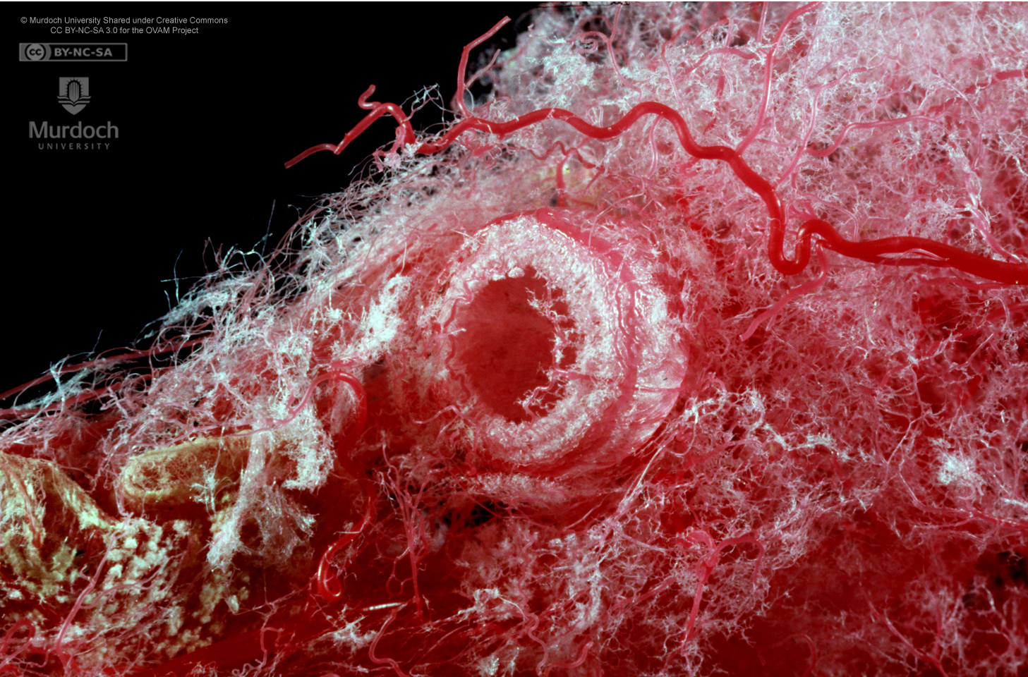
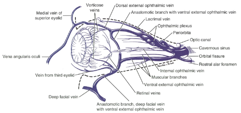
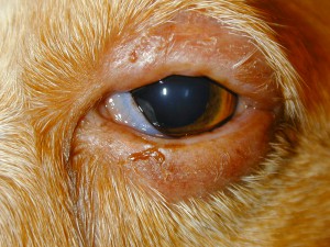
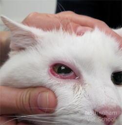
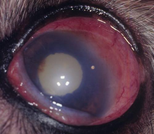
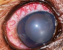
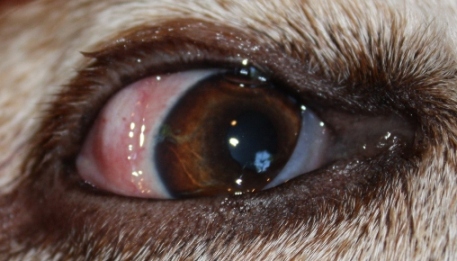
“Red eye” (also called ocular surface hyperemia) is one of the most common eye conditions for which pets are presented to their family veterinarian. Pet parents and some veterinarians occasionally assume reddening of the eyes is just caused by allergies (called allergic conjunctivitis); but the red appearance can be sign of a much more serious and potentially blinding problem, including glaucoma. Understandably the anatomy of the eye, including the vasculature (aka: blood vessels), is quite intricate. For simplicity’s sake, eye blood vessels can be classified as deep (called episcleral) or superficial (called conjunctival). There are two major reasons for these vessels to appear reddened:
- Dilation because of inflammation
- Engorgement because of decreased blood flow (called venous return)
A veterinarian must distinguish which vessels are involved in the reddening process to make an accurate diagnosis and prescribed the most appropriate intervention.
Common causes of superficial reddening include:
- Blepharitis (inflammation of the eyelid)
- Conjuncitivitis (inflammation of the conjunctiva)
- Superficial keratitis (inflammation of the cornea)

Commony causes of deep/episcleral reddening are:
- Deep keratitis
- Uveitis (inflammation of uvea or middle layer of the eye)

- Glaucoma (increased pressure inside the eye)
- Scleritis (inflammation of the outer layer of the eye or “white of the eye”)
- Disease of the orbit (aka: eye socket)
Red eye – essential diagnostic tests
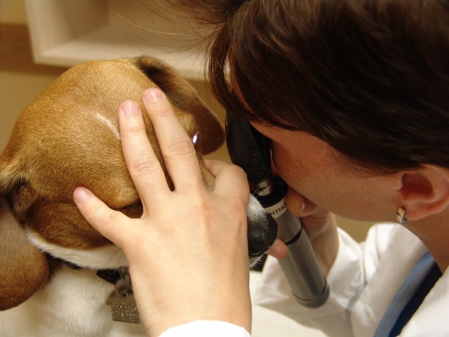
Physical examination: A veterinarian must perform a complete physical examination on any patient with a red eye. Although the eyes are obviously important, it is pivotal an initial evaluation of a patient with a red eye(s) not just focus on the eyes. The rest of the body may hold clues as to what the cause of reddening is (and vice versa)! During the physical examination, you should see the veterinarian use special instruments to evaluate the eyes, including:
- A direct or indirect ophthalmoscope to look in the back of eyes
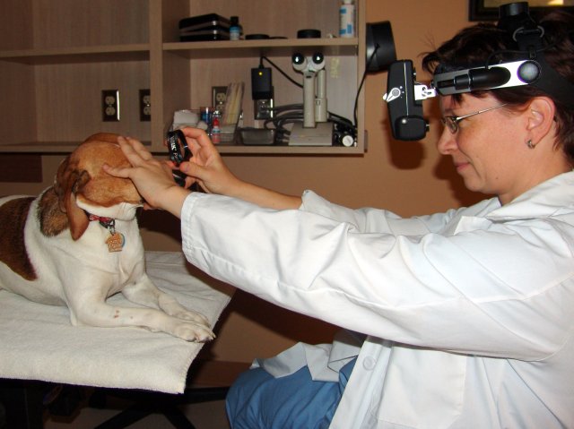
- A biomicroscope (also called a slit lamp) to assess pupil size, shape and equality, as well as to pinpoint lesions on/in specific structures of the eye

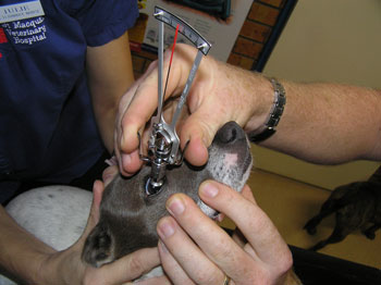
The veterinarian should also apply some gentle direct pressure to each eyelid, attempting to push each eye deeper into its respective orbit/eye socket. This is called retropulsion, and is a technique used to help determine if there is anything causing problems behind the eye itself.
In addition to complete physical and ophthalmic examinations, a few key eye tests are also required to properly and thoroughly evaluate pets with “red eye.” These tests are often called “the big 3” to emphasis their importance, and include:
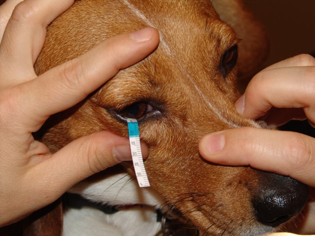
- Schirmer Tear Test: a test to measure the amount of tears a pet can make
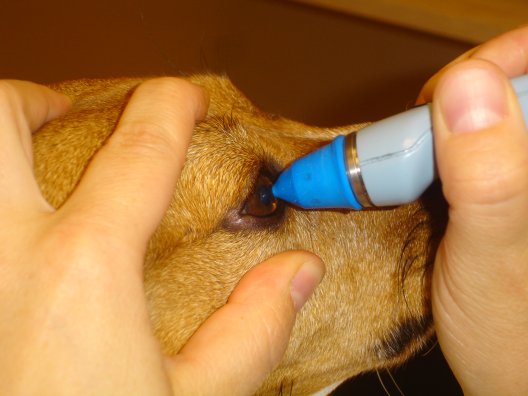
- Tonometry: a test to measure the pressure inside the eyeball (called intraocular pressure; this test is most commonly performed in veterinary medicine via either applanation tonometry (using a Tono-Pen) or indentation tonometry (using a Schiotz tonometer).
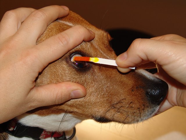
- Fluorescein dye test: a test to check for ulcers on the cornea (the clear part of the eye)
The take-away message about red eye in dogs and cats…
“Red eye” in a dog or cat should raise immediate concerns for pet parents, and they should seek immediate veterinary medical attention for an affected pet. There are many potential causes of “red eye” and accordingly a thorough diagnostic investigation is of paramount importance. Primary care veterinarians are a family’s first line of defense against this clinical problem, and consulting with a board-certified veterinary ophthalmologist is often invaluable to maximize the likelihood of no compromise or loss of vision.
To find a board-certified veterinary ophthalmologist, please visit the American College of Veterinary Ophthalmologists.
To find a board-certified veterinary internal medicine specialist, please visit the American College of Veterinary Internal Medicine.
Wishing you wet-nosed kisses,
cgb