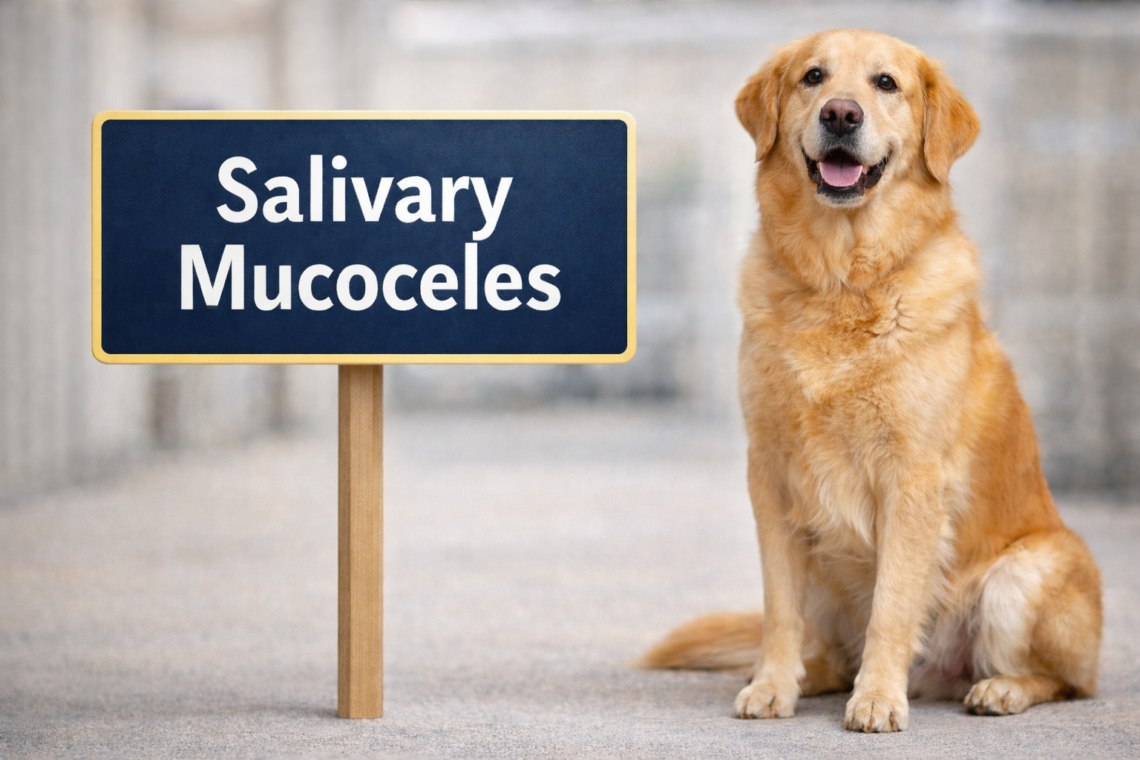

What Is a Salivary Mucocele?
A salivary mucocele is the most common disorder of the salivary glands in dogs. Rather than a true cyst with an epithelial lining, a mucocele represents an extravasation of saliva into surrounding tissues following leakage from a damaged salivary gland or duct. The body reacts to this saliva as an irritant, resulting in a localized collection of thick, mucoid fluid surrounded by inflammatory tissue.
Salivary mucoceles most often involve the mandibular and sublingual salivary glands, though the zygomatic and parotid glands can also be affected. The condition is typically unilateral and can occur in dogs of any age or breed, though certain breeds (e.g., Dachshunds, Poodles, Australian Silky Terriers) may be overrepresented.
Why Do Salivary Mucoceles Develop?
In many patients, a definitive cause is never identified. However, the underlying issue is always disruption of normal salivary flow, allowing saliva to escape into adjacent tissues.
Recognized or suspected causes include:
- Blunt or penetrating trauma (e.g., bite wounds, chewing sticks)
- Foreign bodies within salivary ducts
- Sialoliths (salivary stones), though uncommon in dogs
- Iatrogenic injury (rare; e.g., surgical or dental procedures)
- Idiopathic duct rupture or glandular leakage
Importantly, infection is not the primary driver in most cases, although secondary infection can occur.
Clinical Presentations: Location Dictates Signs
The clinical appearance of a salivary mucocele depends on which gland is affected and where saliva accumulates. Recognizing these patterns is essential for timely diagnosis.
Cervical Mucocele (Most Common)
- Soft, fluctuant, non-painful swelling in the ventral neck
- Typically lateralized, but may appear midline
- Slowly progressive enlargement
Sublingual Mucocele (Ranula)
- Swelling under the tongue on the floor of the mouth
- May be translucent or bluish
- Can interfere with eating or swallowing if large
Pharyngeal Mucocele
- More subtle externally
- Signs of upper airway obstruction:
- Stertor
- Dyspnea
- Dysphagia
Zygomatic Mucocele
- Swelling below the eye
- Exophthalmos (globe protrusion)
- Pain on opening the mouth
A key clinical feature across presentations is that these swellings are often non-painful and compressible, which can delay owner recognition and presentation.
Diagnostic Approach: Confirming the Suspicion
Diagnosis is often straightforward with appropriate clinical suspicion. However, determining the affected gland and excluding other differentials is critical.
Fine-needle aspiration (FNA) is typically the first-line diagnostic step. The aspirated fluid is characteristically:
- Thick, viscous, and stringy
- Clear to blood-tinged
- Low cellularity with mucin and scattered inflammatory cells
Cytology helps rule out abscesses, neoplasia, or lymphadenopathy.
Advanced imaging may be warranted in select cases:
- Ultrasound: Helpful for identifying fluid pockets and guiding aspiration
- CT or MRI: Particularly useful for zygomatic or pharyngeal mucoceles
- Sialography: Rarely used in modern practice
Treatment: Why Surgery Is Best Practice
Definitive treatment requires addressing the source of leakage—not just draining the accumulated saliva.
Surgical excision of the affected salivary gland(s) is the gold standard. For mandibular/sublingual mucoceles, this involves removal of the mandibular-sublingual gland complex, typically via a lateral approach.
For ranulas, a procedure called marsupialization may be performed in addition to gland removal, creating a permanent opening to allow drainage into the oral cavity.
Key practical points:
- Simple drainage alone results in near-universal recurrence
- Surgical success rates are high, with low recurrence when the correct gland complex is removed
- Identifying and removing the ipsilateral gland complex is critical, even if swelling appears midline
Postoperative Care and Prognosis
Most dogs recover uneventfully following surgery. Mild postoperative swelling or seroma formation can occur but is generally self-limiting.
Postoperative management typically includes:
- Analgesia
- Short course of anti-inflammatory therapy
- Monitoring for swelling, discharge, or recurrence
The prognosis is excellent, with recurrence rates reported to be low when surgery is performed correctly.
Complications: What to Watch For
Although uncommon, potential complications include:
- Recurrence (usually due to incomplete gland removal or wrong side identified)
- Damage to adjacent structures (e.g., lingual nerve, hypoglossal nerve)
- Seroma or hematoma formation
- Infection (rare)
Zygomatic mucoceles carry additional surgical complexity due to orbital proximity.
Differential Diagnoses
Several conditions can mimic salivary mucoceles and should be considered:
- Abscess (typically painful, febrile, purulent aspirate)
- Neoplasia (firm, irregular, often progressive)
- Lymphadenopathy
- Cysts or hematomas
FNA is invaluable for distinguishing among these.
Practical Takeaways for the Veterinary Team
- Any non-painful, fluctuant swelling in the neck or oral cavity should prompt consideration of a salivary mucocele
- FNA is diagnostic in most cases and should be performed early
- Do not rely on repeated drainage—this delays definitive care
- Surgical planning must focus on correct gland identification and complete excision
- Client communication should emphasize the benign nature of the condition and the high success rate with surgery
Guidance for Dog Owners
If you notice a soft swelling under your dog’s jaw, tongue, or near the eye, it’s important to have it evaluated promptly. While salivary mucoceles are not cancerous, they do not resolve on their own and can grow large enough to interfere with breathing or eating.
Treatment is highly effective, and most dogs return to normal quickly after surgery. Early evaluation helps ensure a smoother, less complicated recovery.
The Bigger Picture: The Triad of Care
Successful management of salivary mucoceles reflects the strength of the triad of care: the pet owner, the primary care veterinarian, and, when needed, a surgical specialist. Early recognition by owners, prompt diagnosis by the veterinary team, and timely surgical intervention together lead to excellent outcomes.