In previous posts I wrote about variety of urinary tract problems, including tumors of the urinary bladder and protein losing nephropathy (PLN). This week I wanted to share information about another urinary issue – ectopic ureter. I hope you find the post interesting and informative. Happy reading!

Ectopic Ureter – What is it?
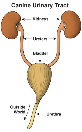
To best understand ectopic ureter, one needs to have a basic understanding of urinary tract anatomy. Dogs and cats have two kidneys. Each kidney is connected to the urinary bladder by a tubular structure called a ureter. The urinary bladder is also connected to the outside world by another tubular structure called the urethra. At the beginning of the urethra are two sphincters or circumferential muscles that constrict to occlude the urethra. Think of these sphincters as doors. When the doors are closed, a pet can’t urinate. The opening and closing of these sphincters are controlled by a complicated system.

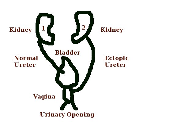
An ectopic ureter is a congenital defect characterized by the ureter connecting to the urinary bladder in an abnormal location. Normally, each ureter enters the urinary bladder before the urethral sphincters. In patients with ureteral ectopia, one or both ureters insert after the urethral sphincters, allowing urine to leak out to cause urinary incontinence.

Ectopic Ureter – What does it look like?
As ectopic ureter is a congenital problem, it is most commonly recognized in pediatric patients; that is, those less than six months of age. Occasionally, however, clinical signs don’t manifest until middle age. There is no sex predisposition, but various breeds are over-represented, including:
- Toy & miniature poodles
- English bulldogs
- Siberian huskies
- Newfoundlands
- Golden retrievers
- Fox terriers
- Labrador retrievers
- Skye terriers
- West Highland white terriers

The primary clinical sign associated with ectopic ureter is urinary incontinence. Some dogs dribble urine as they leak. Many leave “wet spots” on their bed while sleeping. In female dogs, wet and/or discolored fur under the tail and on the inner thighs is not uncommon. Recurrent and/or persistent urinary tract infections are also frequently encountered.
Ectopic Ureter – How is it diagnosed?
Advanced imaging is needed to identify an ectopic ureter. Options include:
- Cystoscopy – the use of special fiber optic camera to look inside the urinary tract while a patient is under anesthesia
- Contrast study –a special dye is injected intravenously and can be followed coursing through kidneys, ureters, urinary bladder, and urethra via radiography (x-rays) or computed tomography (CT scan); the doctor will take series of images in an attempt to identify an ectopic ureter
- Ultrasonography – occasionally an ectopic ureter can be visualized via sonography of the urinary tract
Screening blood and urine tests are recommended to screen for abnormal kidney function and a bacterial urinary tract infection. Pet parents will likely find it helpful to consult with a board-certified veterinary internal medicine specialist to develop a logical and cost-effective diagnostic plan.
Ectopic Ureter – How is it treated?
Ectopic ureter is most commonly corrected either via a laser technique or surgery. Both procedures require extensive training and specialized equipment. A laser can be used in conjunction with cystoscopy to redirect the opening of the ureter so it’s located before the urethral sphincters. See the video below to see this procedure performed by a board-certified veterinary surgeon.
Surgery involves physically moving the ureter to a different location so it enters the urinary bladder at a more appropriate anatomic location – this procedure is called a neoureterocystostomy. Families are recommended to partner with a board-certified veterinary surgeon to help determine the best therapeutic course of action. The prognosis for both procedures is good, and the majority of dogs don’t have urinary incontinence long-term. Patients with residual incontinence are usually able to be adequately controlled with medication. Those with chronic urinary incontinence after laser or surgical intervention after have involvement of the urethral sphincters.
The take-away message about ectopic ureter in dogs…
Ectopic ureter is a congenital defect that causes urinary incontinence in dogs. Those affected are typically younger than six months of age, and certain breeds of over-represented. Advanced imaging is required for definitive diagnosis. With laser or surgical intervention, the majority of dogs have a good prognosis.
To find a board-certified veterinary internal medicine specialist, please visit the American College of Veterinary Internal Medicine.
To find a board-certified veterinary surgeon, please visit the American College of Veterinary Surgeons.
Wishing you wet-nosed kisses,
CriticalCareDVM


