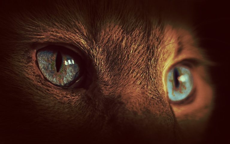
Those who regularly read my blog (I hope that’s most of you!) know how much I love eyes. Those colorful globes are simply a site to behold! Although cliché, I believe eyes truly are the windows to the soul. This week I wanted to share information about a unique eye condition that can occur in dogs and cats – Horner’s Syndrome. I hope you enjoy it and find the content shareworthy. Happy reading!
Horner’s Syndrome – What is it?
The eyes and surrounding structures have many intricate nerves controlling their functions and movements. Horner’s syndrome results from a dysfunction of the sympathetic nerves that feed the eyes. The sympathetic nervous system is part of the autonomic nervous system, and is often called the “fight or flight” system. When it comes to the eyes, the sympathetic nerves keep:
- Eyes in the front of the socket
- Nictitans (3rd eyelid) recessed
- Upper eyelids fully raised
- Pupils appropriately open
When the sympathetic nerves to the eyes aren’t functioning properly, the above-listed anatomic functions don’t happen. We will review these changes in more detail in just a little bit.
We often are unable determine the cause of Horner’s syndrome in dogs or cats. Indeed, the reason is not identifiable in ~50% of all patients. Thus, we use the term idiopathic to describe Horner’s Syndrome in these patients. As you may know, idiopathic is simply the medical way of saying, “We don’t know.”
To call a disease process idiopathic, one must first rule out any other potential identifiable cause, including:
- Trauma – Injuries to cervical and thoracic spinal cord can result in unilateral and bilateral Horner’s Syndrome
- Lesions in the chest cavity – Masses and lymph node enlargement at the level of the thoracic inlet or in the cranial mediastinum may induce Horner’s syndrome if they affect the sympathetic nerves in these areas
- Inner ear disease – Infection, inflammation, and/or tumors in this part of the body can affect the sympathetic nerves to induce Horner’s Syndrome
- Lesions behind the eyes – Abscesses, tumors, and/or injury to the area behind the globes of the eye (called the retrobulbar space) have been associated with the development of Horner’s Syndrome
- Diabetes mellitus – Horner’s Syndrome due to nerve dysfunction induced by diabetes mellitus has been documented in dogs
Horner’s Syndrome – What does it look like?
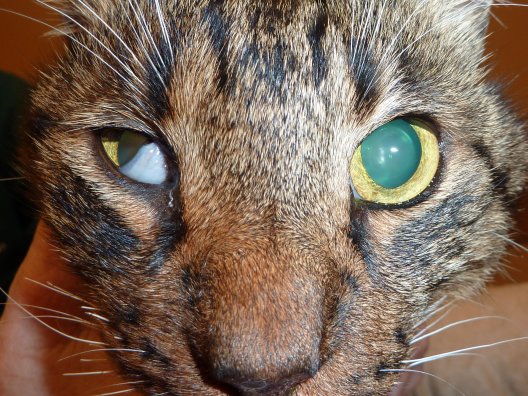
Horner’s Syndrome may affect one or both eyes. There is no breed, age, or sex predilection. Patients with Horner’s Syndrome have four classic changes to their eye(s):
- Miosis – Sympathetic dysfunction causes the affected pupil(s) to constrict
- Protrusion of the nictitans – Without properly functioning sympathetic nerves, the 3rd eyelid doesn’t fully retract
- Ptosis – When the sympathetic nerve going to the superior tarsal muscle (also called Muller’s muscle) isn’t working properly, a pet can’t open its upper eyelid fully
- Enophthalmos – With dysfunctional sympathetic nerves, the muscles of the orbits retract slightly causing the globe to appear sunken in the orbit
Other potential signs affiliated with Horner’s Syndrome are dilation of the skin blood vessels and subsequently pink appearance to the skin of the ears. Mild nasal congestion may also be noted, but these signs are often difficult to detect.
Horner’s Syndrome – How is it diagnosed?
Diagnosing Horner’s Syndrome is relatively straightforward. Observed a constricted pupil, incompletely raised upper eyelid, sunken eye, and elevated 3rd eyelid is diagnostic. Once diagnosed, the veterinarian should begin a logical approach to trying to identify an underlying cause. After performing a complete physical examination, the veterinarian will recommend performing some specific tests:
- Complete blood count – a non-invasive blood test to evaluate red blood cells, white blood cells, and platelets
- Serum biochemical profile – a non-invasive blood test to assess liver and kidney function, as well as electrolytes (i.e.: sodium, potassium) and some gastrointestinal enzymes
- Urinalysis – a non-invasive urine test to evaluate kidney function and screen for inflammation and infection in the urinary tract
- Chest radiographs (x-rays) – non-invasive imaging test to look for evidence of masses in the chest cavity
- Advanced imaging – some patients benefit from computed tomography (CT scan) and magnetic resonance imaging (MRI)
Sympathetic nerves to the eye are relatively long, and are composed of three neurons. A neuron is simply a nerve cell that transmits nerve impulses. The first neuron starts in the hypothalamus in the brain and ends in the first part of the thoracic spinal cord. The second neuron starts in this location, follows a unique pathway out of the spinal cord into the sympathetic trunk and ends in an area called the cranial cervical ganglion. As the second and third neurons meet at the cranial cervical ganglion, non-invasive pharmacologic testing can be performed to determine where along the course of the nerve is the lesion. Lesions involving the second neuron are termed preganglionic and those affecting the third neuron are described as postglanglionic.

Horner’s Syndrome – How it is treated?
There is no specific treatment for idiopathic Horner’s Syndrome. Efforts should be made to determine any possible underlying cause, and then treat any identified disease accordingly. Preganglionic lesions have a less favorable prognosis than postganglionic ones. Approximately 50% of those with idiopathic disease improve or resolve within 6-8 weeks. Thankfully, Horner’s Syndrome is not painful, and patients are not away of the noted eye changes.
The take-away message about Horner’s Syndrome in dogs and cats…
Horner’s Syndrome is an ophthalmic syndrome in dogs and cats characterized by specific eye changes. Diagnosis is relatively straightforward, but determining the underlying cause is often unrewarding. Nevertheless, a thorough diagnostic investigation is needed to ensure there is identifiable cause. There is no specific therapy for Horner’s Syndrome, and this condition does not cause discomfort in affected pets.
To find a board-certified veterinary ophthalmologist, please visit the American College of Veterinary Ophthalmologists.
Wishing you wet-nosed kisses,
cgb


