
I’ve said it before. I love eyes…albeit not enough to want to be an ophthalmologist, but at least enough to respect their function and beauty. Unfortunately, sometimes our pets develop serious eye conditions. This week’s post is dedicated to a specific intraocular (inside the eye) condition called hyphema. I hope this information is interesting to you. Happy reading!
What is hyphema?
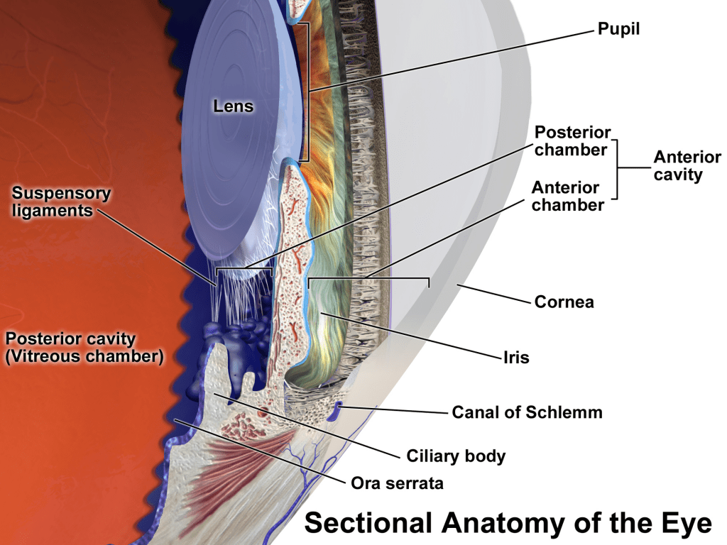
To understand hyphema, one must have a basic understanding of eye anatomy. The clear part of the eye is called the cornea. The colored part is the iris, the borders of which form the pupil. The area between the cornea and the iris is called the anterior chamber. Behind the iris is the posterior chamber and the lens. Behind the lens is the vitreous chamber and retina.
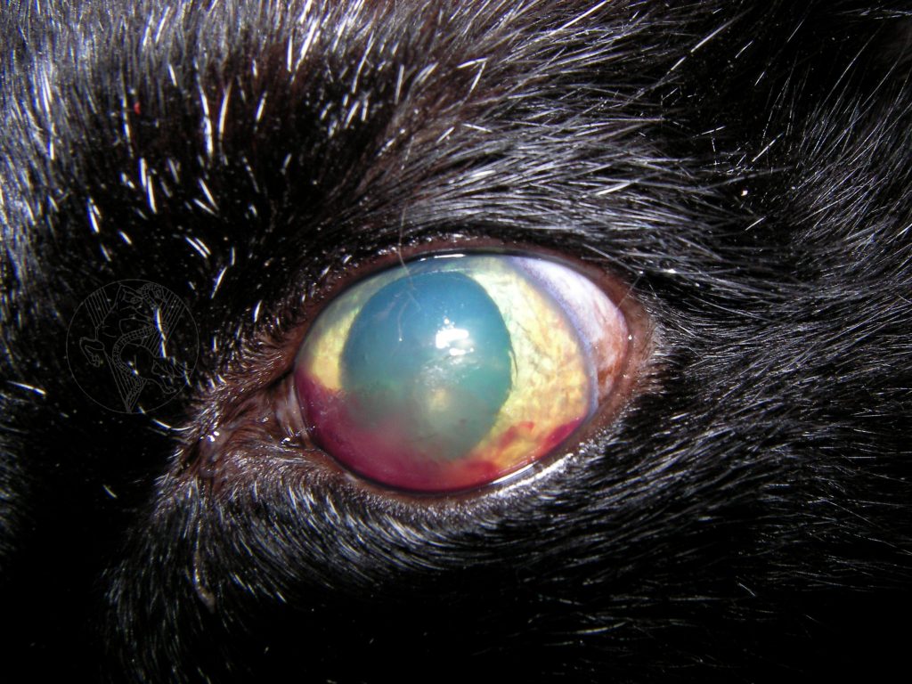
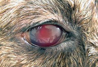
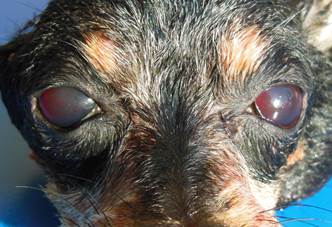
Hyphema is bleeding into the anterior chamber of the eye and is usually associated with a breakdown of the blood ocular barrier and subsequent inflammation (called uveitis). Common causes of hyphema in dogs and cats are:
- Ocular trauma
- Retinal detachment
- Uveitis
- Cancer (both intraocular & systemic)
- Congenital & heritable eye disorders (i.e.: vitreoretinal dysplasia in Labrador retrievers; rupture of a persistent hyaloid artery)
- Clotting disorders
- Hypertension
- Thromboembolic disease
- Hyperviscosity syndromes
- Thrombocytopenia (low platelets)
- Infectious diseases (i.e.: leptospirosis, Rocky Mountain spotted fever, feline infectious peritonitis)
What does it look like?
The degree of hemorrhage in the anterior chamber is variable. Sometimes both eyes are effected. Sometimes only one. Sometimes there’s bleeding in other parts of the body. The type and extent of hyphema depends on the underlying cause.
How is hyphema diagnosed?
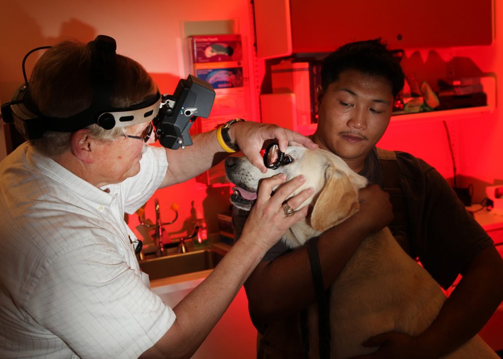
Diagnosing hyphema is straightforward. One can typically readily visualize blood in the anterior segment of the eye. The challenging aspect of hyphema is figuring out what caused it. Veterinarians will obtain a thorough patient history and perform a complete physical examination. They will subsequently recommend an appropriate diagnostic investigation that may include:
- Non-invasive eye tests – Schirmer tear testing, flourescein staining, tonometry
- Complete blood count
- Biochemical profile
- Urinalysis
- Coagulation profile
- Infectious disease screening (extent/type dependent on geographical area)
- Diagnostic imaging (i.e.: ocular ultrasound, chest radiographs/x-rays, abdominal sonography)
- Blood pressure measurement
Pet owners may find it helpful to partner with a board-certified veterinary ophthalmologist to develop a logical and cost-effective diagnostic plan.
How is hyphema treated?
Effective treatment of hyphema is dependent on accurately identifying the underlying cause. For this reason, a thorough diagnostic investigation as described earlier is of paramount importance.
Some patients can be effectively managed with ophthalmic medications. Some need systemic therapies, including antibiotics and anti-inflammatory drugs. Others benefit from surgery. Patients with coagulopathies often benefit from plasma infusions. Pet owners are encouraged to partner with their family veterinarian and a board-certified veterinary ophthalmologist
The take-away message about hyphema in dogs & cats…
Hyphema is an important eye condition characterized by bleeding in the anterior chamber of the eyes. There are a myriad of potential causes, and thus making an accurate diagnosis is essential for initiating the most appropriate therapies.
To find a board-certified veterinary ophthalmologist, please visit the American College of Veterinary Ophthalmologists.
Wishing you wet-nosed kisses,
CriticalCareDVM