
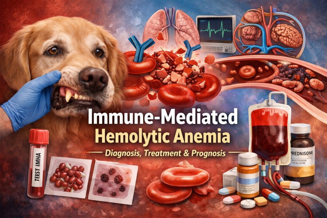

Immune-mediated hemolytic anemia (IMHA) is one of the most common life-threatening causes of anemia in dogs and remains an important, though less frequent, disease in cats. In many patients, IMHA presents as a true medical emergency. Therefore, rapid recognition, accurate diagnosis, and timely intervention are essential for survival.
What Is Immune-Mediated Hemolytic Anemia (IMHA)?
IMHA occurs when a dog’s or cat’s immune system mistakenly targets and destroys its own red blood cells. As red blood cell destruction accelerates beyond the bone marrow’s ability to compensate, anemia develops. Consequently, oxygen delivery to tissues declines and may result in organ dysfunction or death.
Veterinarians classify IMHA into two major categories:
Non-Associative (Primary or Idiopathic) IMHA
- No identifiable underlying trigger
Associative (Secondary) IMHA
- Immune-mediated hemolysis occurs in association with:
- Infectious disease (for example, tick-borne disease)
- Neoplasia
- Inflammatory or immune-mediated disorders
- Drug exposure or immune stimulation
Importantly, identifying whether IMHA is non-associative or associative directly influences treatment selection, prognosis, and duration of immunosuppressive therapy.
Why Diagnosing IMHA Is Challenging
One of the most important messages from the ACVIM consensus statement is clear:
No single test definitively diagnoses IMHA in all patients.
Instead, IMHA is a clinicopathologic diagnosis that requires combining multiple findings. Over-reliance on any single test, such as a Coombs’ test alone, increases the risk of misdiagnosis.
To diagnose IMHA with confidence, veterinarians must demonstrate:
- Anemia
- Immune-mediated red blood cell destruction (hemolysis)
Step 1: Confirming Anemia and Hemolysis
Confirming Anemia
A complete blood count (CBC) confirms anemia. In addition, affected dogs and cats often show:
- Lethargy or weakness
- Pale mucous membranes
- Tachycardia or tachypnea
- Collapse in severe cases
Evidence of Hemolysis
Hemolysis indicates active red blood cell destruction and may present as:
- Icterus (jaundice) of the gums, sclera, or skin
- Hyperbilirubinemia on serum biochemistry
- Hemoglobinemia or hemoglobinuria
Although these findings confirm red blood cell destruction, they do not independently confirm immune-mediated disease.
Step 2: Demonstrating Immune-Mediated Red Blood Cell Destruction
The ACVIM consensus recommends using multiple immune-based tests whenever possible.
Saline Agglutination Testing
- Detects antibody-mediated red blood cell clumping
- A strong positive result strongly supports IMHA
- Improper technique may produce false-positive results
Direct Antiglobulin (Coombs’) Test
- Detects antibodies or complement bound to red blood cells
- A positive result supports IMHA
- A negative result does not exclude IMHA, particularly early or previously treated cases
Blood Smear Evaluation
- Spherocytes are a classic finding in dogs
- Smear review also helps rule out alternative causes of anemia, such as parasites or red blood cell fragmentation
Flow Cytometry (When Available)
- Detects immunoglobulins on red blood cell membranes
- Useful in select cases, although not widely available
Key takeaway:
The ACVIM consensus recommends identifying at least two indicators of immune-mediated red blood cell destruction, combined with evidence of hemolysis, before confidently diagnosing IMHA.
Why Screening for Underlying Causes Matters
Before diagnosing non-associative IMHA, clinicians should actively evaluate for secondary triggers, including:
- Infectious disease
- Neoplasia
- Inflammatory conditions
- Drug exposure
This step is critical because:
- Treating the underlying disease may reduce immune activation
- Prognosis differs between primary and secondary IMHA
- Some patients require less aggressive or shorter-term immunosuppression
For pet owners, this explains why additional testing is often recommended even after IMHA is suspected.
Treating Immune-Mediated Hemolytic Anemia
Immunosuppression Is the Cornerstone of Therapy
Most dogs with IMHA require immunosuppressive medications to stop ongoing red blood cell destruction. Corticosteroids, such as prednisone, form the foundation of treatment. In addition, current guidelines support adding a second immunosuppressive agent early to improve outcomes.
Common options include:
- Cyclosporine
- Mycophenolate
- Azathioprine (dogs only)
- Leflunomide
As a result, treatment often involves multiple medications, frequent monitoring, and gradual dose adjustments over weeks to months.
Blood Transfusions Can Be Life-Saving
Many dogs with IMHA develop severe anemia and require blood transfusions for stabilization. Importantly, the need for transfusion does not indicate treatment failure. Instead, transfusions provide essential support while immunosuppressive medications take effect.
Blood Clots Are a Major Risk
Dogs with IMHA are at high risk for thrombosis affecting the lungs, brain, or other vital organs. Therefore, thromboprophylaxis is a critical component of therapy.
The consensus guidelines support antithrombotic medications, such as clopidogrel, with drug selection tailored to each patient’s risk factors and clinical status.
Not Every Therapy Works for Every Dog
Adjunctive therapies, including intravenous immunoglobulin (IVIG), splenectomy, or therapeutic plasma exchange, may benefit select patients. However, evidence remains mixed, and these treatments are not universally recommended.
This variability highlights an essential principle: IMHA treatment must be individualized, and response to therapy varies widely.
What This Means for Pet Owners
Treatment for IMHA is often intensive and may require hospitalization, frequent rechecks, and long-term medication. Recovery can be slow, and relapses may occur. Nevertheless, many dogs survive and enjoy a good quality of life.
Ask your veterinarian or a board-certified specialist why specific treatments are recommended and what monitoring is required. Managing IMHA is a team effort.
The Bottom Line
The ACVIM consensus guidelines represent a major step forward in standardizing care for canine IMHA. By combining evidence-based medicine with expert clinical judgment, these recommendations aim to improve outcomes while acknowledging the complexity of this disease.


Great column – look forward to sharing it with clients who have pets with this hard-to-understand condition!
Thanks for reading & sharing! I appreciate the support!
How is this different from aplastic anemia? The treatment seems to be the same.
True aplastic anemia is characterized by decreased production of red blood cells (and white blood cells and platelets) by the bone marrow which results in decreases in all cell lines (called pancytopenia). Normal blood cell producing tissue is replaced by fat. Aplastic anemia can be divided into acute or chronic forms; both involve decreased proliferation or destruction of blood stem cells in the marrow. With the acute form, a patient usually first has low white blood cells and then low platelets. As red blood cells “live” longer than white blood cells and platelets, anemia does not become apparent until the later stages. Acute aplastic anemia is potentially reversible if the initiating cause is eliminated. In comparison, the prognosis for the chronic form is less favorable and marrow recovery, if it occurs, may take weeks to months.
Thank you for your reply! It was very helpful!
I’m glad you found the information helpful!
We are a group of volunteers and opening a new scheme in our community. Your site offered us with valuable info to work on. You’ve done a formidable job and our whole community will be thankful to you.