
Introduction
Tracheostomy is a critical and sometimes lifesaving procedure in both dogs and cats. Whether performed emergently in response to upper airway obstruction or electively as part of planned postoperative airway management, the procedure demands careful patient selection, meticulous technique, and vigilant postoperative care. Understanding when and how to perform a tracheostomy and recognizing potential complications can significantly improve patient outcomes in both general and referral practice settings.
Indications for Tracheostomy
Perform a tracheostomy when significant upper airway obstruction prevents adequate airflow through the larynx, pharynx, or nasal passages and cannot be rapidly relieved by other means. Common indications include:
- Laryngeal paralysis (bilateral)
- Brachycephalic obstructive airway syndrome (BOAS) with dynamic collapse
- Neoplasia affecting the larynx, pharynx, or nasal cavity
- Severe upper airway trauma (bite wounds, fractures, thermal injury)
- Foreign body obstruction that cannot be removed immediately
- Postoperative laryngeal or pharyngeal swelling (e.g., following upper airway surgery)
The decision to perform a tracheostomy should always consider whether the obstruction is likely to resolve or persist. If the cause is transient (e.g., postoperative swelling), a temporary tracheostomy may suffice. A permanent tracheostomy may be indicated for irreversible or progressive diseases (e.g., neoplasia, end-stage laryngeal collapse),
Emergency vs. Elective Tracheostomy
Emergency Tracheostomy
Perform an emergency tracheostomy when an airway obstruction prevents effective oxygenation and intubation is not possible. These situations often occur with acute upper airway obstruction or severe laryngeal edema. The goals are rapid airway access and oxygenation.
Key features:
- Performed under minimal or no anesthesia due to severe hypoxemia.
- Quickly make a vertical skin incision over the ventral midline, typically between the cricoid cartilage and the 4th tracheal ring.
- The area between two tracheal rings are incised transversely to create an opening.
- A tracheostomy tube (or, in dire cases, an appropriately sized endotracheal tube) is placed immediately.
- The airway is secured and ventilation restored before additional stabilization or closure steps.
This is a time-critical, high-stress procedure where speed and oxygenation take priority over perfect surgical finesse.
Elective Tracheostomy
An elective tracheostomy is performed in a controlled setting when upper airway obstruction is anticipated, such as during recovery from upper airway surgery or as part of a staged plan for chronic airway disease.
Key features:
- Performed under general anesthesia with full aseptic preparation.
- The vertical skin incision is centered over the mid-cervical trachea (typically 3rd–5th tracheal rings).
- A horizontal incision is made through two tracheal rings, avoiding damage to adjacent cartilage and the trachealis muscle.
- Stay sutures are placed around the cranial and caudal tracheal rings to facilitate tube replacement.
- A sterile tracheostomy tube (often a double-cannula design for large dogs or single-cannula for cats and small dogs) is inserted and secured with umbilical tape or sutures around the neck.
- The skin may be loosely apposed around the tube to minimize subcutaneous emphysema.
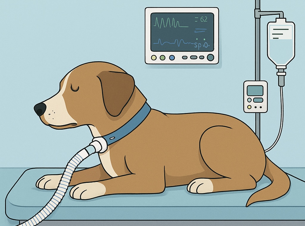
Postoperative Care and Tube Management
Effective postoperative care is critical for success. Veterinary technicians play a central role in maintaining tube patency and patient safety.
Key aspects of care:
- Continuous monitoring: Watch for increased respiratory effort, stertor, or bubbling around the stoma.
- Tube patency: Suction as needed using sterile technique; humidify inspired air to prevent crusting.
- Tube cleaning: Remove and clean or replace the inner cannula every 1–2 hours initially, then as needed.
- Stoma hygiene: Gently clean with saline to remove discharge and prevent infection.
- Prevent occlusion: Avoid bedding or bandaging material near the stoma; ensure the patient cannot rub the site.
- Humidification: Use nebulization or humidified oxygen to maintain mucociliary clearance.
- Oxygen supplementation: Provide as needed based on pulse oximetry and arterial blood gas monitoring.
For temporary tracheostomies, tubes can typically be removed once the primary airway obstruction resolves and the patient tolerates breathing through the upper airway.
Potential Complications
Despite its life-saving potential, tracheostomy carries several potentially serious complications, particularly if tube care is inadequate:
| Category | Examples / Consequences |
|---|---|
| Obstruction | Mucus plugging, kinking, dried secretions — can lead to acute respiratory distress |
| Infection / Inflammation | Stoma cellulitis, tracheitis, pneumonia |
| Mechanical Trauma | Tracheal cartilage necrosis, subcutaneous emphysema, pneumomediastinum |
| Tube displacement | Accidental decannulation or migration, especially in cats |
| Aspiration | Due to bypassing of normal laryngeal reflexes |
| Granulation tissue | Chronic irritation or infection leading to obstruction or stenosis |
Meticulous nursing care and frequent reassessment minimize these risks.
Summary
Tracheostomy, whether emergency or elective, remains a cornerstone intervention in the management of severe upper airway obstruction in dogs and cats. Success depends as much on indication recognition and technical precision as on ongoing postoperative management. Emergency tracheostomies save lives, while elective tracheostomies provide planned airway control when upper airway compromise is expected. By anticipating complications and engaging the entire veterinary team in vigilant postoperative care, clinicians can ensure the best possible outcomes for their patients.