
Wound care is a routine part of small animal practice. Search engines show strong interest in topics like dog wound healing stages, cat wound care at home, bandage types, and Manuka honey for wounds. This post reviews the phases of wound healing, the types of dressings and bandages available, the clinical use of medical grade manuka honey, and the importance of always covering wounds and drains.
Phases of Wound Healing in Dogs and Cats
Understanding the phases of healing helps you choose the right treatment and the right bandage for every patient. Wounds progress through overlapping stages influenced by tissue damage, contamination, patient health, and clinical technique.
Inflammatory phase
• Begins immediately after injury.
• Platelets form a clot and start hemostasis.
• Neutrophils and macrophages remove debris and bacteria.
• Signs include redness, heat, swelling, and exudate.
• Aim to limit contamination, remove necrotic tissue, and support a clean wound bed.
Repair or proliferative phase
• Involves fibroblast activity, angiogenesis, granulation tissue, and epithelialization.
• New collagen forms and wound contraction begins.
• Moist wound healing speeds this phase.
• A clean, moist, protected wound environment is the priority.
Maturation or remodeling phase
• Collagen reorganizes and strength increases.
• Scar tissue forms but never regains full tensile strength.
• Protect the wound from trauma during this period.
Bandages and Dressings Used in Small Animal Wound Care
Choosing the correct bandage is essential for controlling moisture, protecting tissue, directing exudate, and supporting healing. The primary, secondary, and tertiary layers each serve a specific purpose.
Primary contact layer
• Sits directly on the wound.
• Can be non adherent, absorbent, or moisture donating.
• Includes hydrogels, alginates, non adherent gauze, silver impregnated dressings, and honey based dressings.
• Maintain moisture when appropriate or absorb excess exudate.
Secondary layer
• Provides absorption and padding.
• Common materials include rolled cotton and cast padding.
• Controls fluid buildup and maintains an even surface.
Tertiary layer
• Protects the bandage and keeps layers in place.
• Examples include cohesive wrap, elastic tape, and protective coverings.
• Should be snug but not tight enough to affect circulation.
Specialty wound dressings
• Negative pressure wound therapy.
• Medical grade honey dressings.
• Antimicrobial dressings for contaminated or infected wounds.
• Moisture managing foams and hydrocolloids.
Use of Manuka Honey in Veterinary Wound Management
Medical grade Manuka honey is popular in small animal medicine because of its osmotic activity, low pH, and antimicrobial effects. It can help reduce bacterial load, support autolytic debridement, and maintain moisture.
Considerations
• Use only medical grade honey.
• Ideal for contaminated wounds, second intention healing, and wounds that need debridement support.
• Some studies show variable cosmetic outcomes in primary closure incisions, so choose cases carefully.
• Apply as an impregnated dressing or thin layer under the primary bandage.
• Continue to monitor for excessive moisture or maceration.
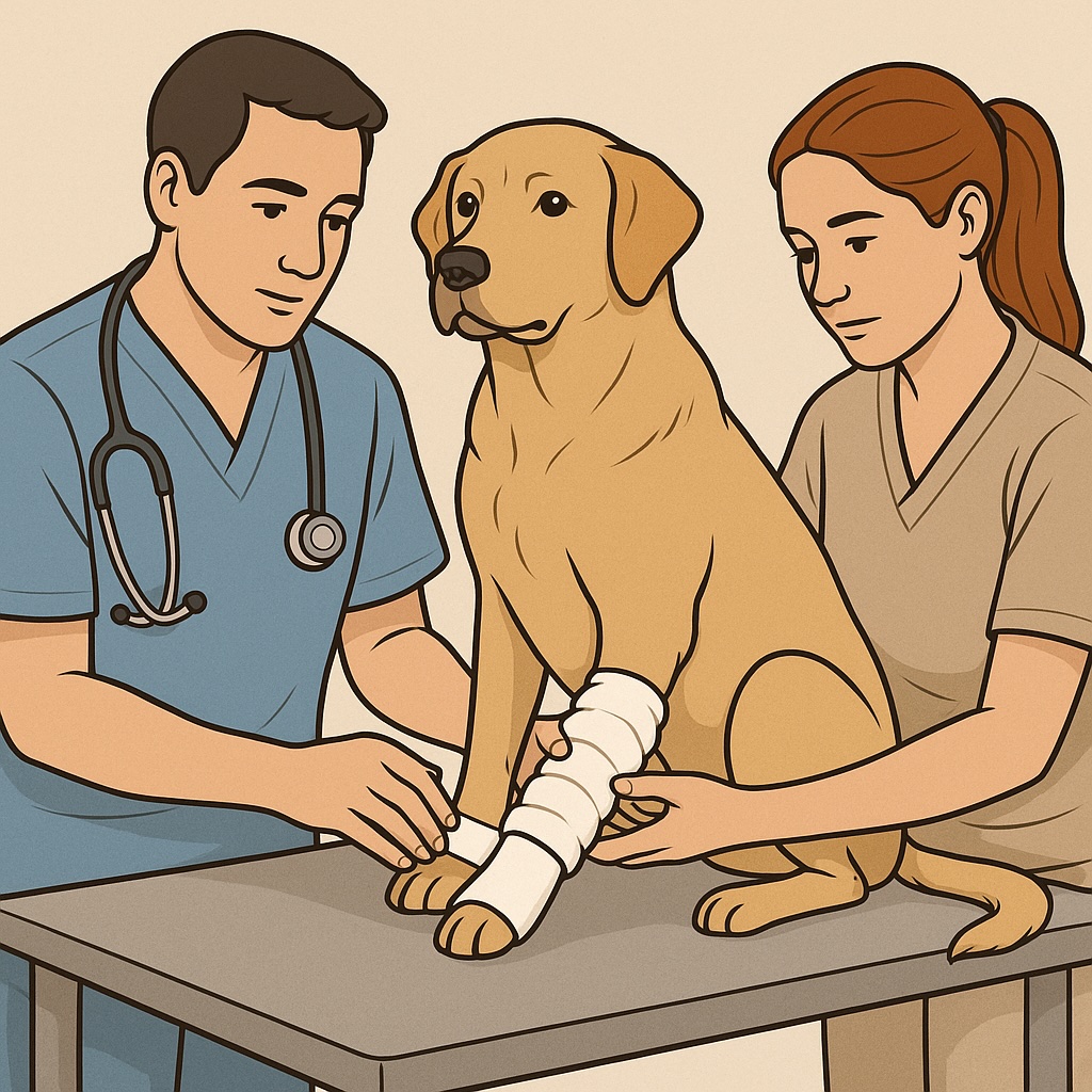
Why All Wounds and Drains Must Be Covered
Covering wounds and drains improves outcomes and reduces complications. Uncovered wounds dry out, attract contamination, and encourage patient interference.
Key reasons to cover every wound
• Protects against environmental contamination.
• Reduces risk of licking, chewing, or scratching.
• Maintains a controlled moisture level that supports the proliferative phase.
• Helps manage exudate and odor.
• Supports secure placement of drains and permits accurate assessment of drainage.
• Allows consistent monitoring of odor, discharge color, and tissue quality.
Drain site management
• Cover the drain exit site with absorbent material.
• Reinforce the area to prevent motion or pulling.
• Keep the entire region covered until the drain is removed.
• Educate owners to keep the bandage dry and return for scheduled rechecks.
Clinical Workflow for Wound Management
Step 1. Initial evaluation
• Stabilize the patient.
• Clip and clean the area around the wound.
• Lavage thoroughly.
• Decide if primary closure or open wound management is appropriate.
Step 2. Debridement and contamination control
• Use mechanical, enzymatic, or surgical debridement as needed.
• Select a primary dressing suited to exudate level.
• Stabilize with a full bandage system.
Step 3. Support the proliferative phase
• Choose moisture maintaining dressings.
• Reassess wound edges, granulation tissue, and drainage.
• Transition dressing type as exudate decreases.
Step 4. Protect during maturation
• Reduce bulk of the tertiary layer once stable.
• Maintain coverage until tissue strength is adequate.
• Schedule regular rechecks.
Key Takeaways for Veterinary Teams
• Understand the phases of healing to direct treatment.
• Select dressings based on exudate, contamination level, and wound age.
• Use medical grade Manuka honey as an adjunct for specific wound types.
• Always cover wounds and drains to protect tissue, manage exudate, and reduce complications.
• Educate clients about bandage care and the need for scheduled rechecks.