Many pet parents have heard of cruciate ligament injuries in pets. Yet few know about another common knee disorder afflicting our furry companions – luxating patellas. The patella, more commonly known as the knee cap, can slip out of its normal groove (aka dislocate or luxate) to cause lameness, discomfort, and inflammation. This week I dedicate some time to this orthopedic condition in an effort to increase awareness of it. Happy reading!
Luxating Patellas – What are they?
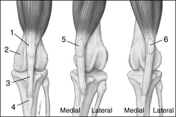
The patella is small bone incorporated in the tendon of the quadriceps muscles of the thigh that attaches to the top of the tibia or shin bone. The combination of the quadriceps muscle, patella, and the tendon are responsible for extending the knee joint. A luxation occurs when the patella moves out of its normal position, and it can move to inside (medial) or outside (lateral) aspect of the knee.

Patellar luxations predominantly affect small dogs, and some are over-representing:
- Yorkshire Terriers
- Pomeranians

- Chihuahuas
- Boston Terriers
- Miniature Poodles
- Shar Peis
- Akitas
- Great Pyrenees
The definitive cause of luxation in all pets in not always known. Certainly a traumatic injury to the knee can result in a dislocation. Many affected dogs have shallow femoral grooves, the boney slots in which knee caps normally reside, predisposing patellas to slip out of place. There is also mounting evidence malformations in other components of the extensor apparatus of the knee contribute to patellar luxations, including:
- Abnormal formation of the shin bone (tibia)
- Abnormal hip joint formation (i.e. hip dysplasia)
- Tightness of the quadriceps muscles
- Elongated patellar ligament
- Malformation of the thigh bone (femur)
Approximately 50% of patients have both of their knees caps affected. Given the cumulative evidence strongly supporting a genetic component to this disease, affected pets should not be bred.
Luxating Patellas – What are the clinical signs?
Many affected pets don’t have any clinical signs of a luxating patella. Often times a veterinarian discovers the problem during a preventative healthcare examination. At times one may observe a pet limp on the affected leg or even hiked it up for a few steps. In severe cases, those with knees caps that luxate to the inside of the knee (medial luxation) appear to have a bow-legged appearance while those that luxate to the outside of the knee (lateral luxation) are described as having a knocked-in knee appearance.
Luxating Patellas – How are they diagnosed?
Definitive diagnosis of a luxating patella is relatively straightforward. A veterinarian is often able to make an accurate diagnosis through simple palpation of the knee in an awake pet. However, some pets do benefit from sedation for this examination. Patellar luxations are graded based on the degree of dislocation:
- Grade I: the patella can be moved out of its normal location, but spontaneously moves back to normal position
- Grade II: the patella spontaneously and intermittently moves out its normal location, and can be replaced back into its normal position
- Grade III: the patella is out of its normal position most of the time, but can be replaced back into its normal position
- Grade IV: the patella is always out of its normal location and can’t be replaced backed in its normal groove
As mentioned earlier, anatomical abnormalities in other components of the knee extensor apparatus are relatively common, and some non-invasive and minimally invasive diagnostic tests are helpful to identify them, including:
- Radiographs/x-rays of the knees, hips, thigh bones (femurs), and shin bones (tibias)
- CT scan of the pelvic/rear/hind legs
- Assessment of the knee for ligamentous damage
Consultation with a board-certified veterinary surgeon can be invaluable to develop a logical and cost-efficient plan for a proper diagnostic investigation.
Luxating Patellas – How are they treated?
Remember not every patient with a patellar luxation has clinical signs. These patient do not typically require surgical intervention, but rather should be serially monitored by their family and family veterinarian. Those with grade II or higher luxations commonly benefit form surgery, and there are several strategies to correct a lunation, including:
- Realignment of the knee extensor apparatus
- Trochleoplasty (deepening of the femoral groove)
- Adjustment of the tension in the soft tissues around the knee joint
Many primary care veterinarians are comfortable performing some of the corrective surgeries.

Other techniques and procedures are quite intricate and require special equipment. Partnering with a board-certified veterinary surgeon can empower pet parents with the information they need to make the best decision for their fur babies. Following surgery, recurrence of patellar instability is uncommon, and the overwhelming majority of pet parents are happy with their pet’s quality of life.
The take-away message about luxating patellas…
Dogs may occasionally be born with knee caps (patellas) that slip out of place or luxate. This can be painful, resulting in tremendous inflammation. Pet parents are encouraged to work with family veterinarians and board-certified veterinary surgeons to develop the best treatment plan, potentially including surgery. With surgery, fur babies with luxating patellas can readily return to comfortable and happy lifestyles.
To find a board-certified veterinary surgeon, please visit the American College of Veterinary Surgeons.
Wishing you wet-nosed kisses,
cgb


