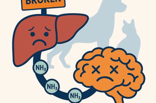
Many dog owners know a finding of elevated blood calcium can be quite concerning. Cancer, including lymphoma and apocrine gland carcinoma of the anal gland, is a leading cause of blood calcium elevations in dogs. Yet, sometimes this elevation is not caused by lymphoma or anal gland cancer! Sometimes, the parathyroid glands in the neck are simply working too hard. This condition is called primary hyperparathyroidism, and this week I’m sharing some important details about this disease. Happy reading!

Primary hyperparathyroidism – What is it?
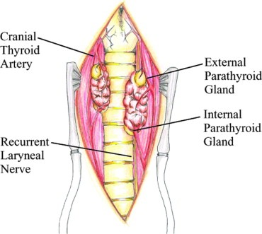
The parathyroid glands are closely associated with the thyroid glands in the neck. Most dogs have four glands, and they are located on or in the capsule of the thyroid gland. Occasionally, parathyroid tissue can be found is other locations in the body, including the mediastinum in the chest cavity.

The parathyroid glands produce, store, and secrete a unique hormone called parathyroid hormone (also called PTH). When blood calcium level decreases, PTH is secreted into the blood to raise the calcium level via some intricate pathways, including:
- Increased resorption of calcium from the kidneys
- Mobilization of calcium from bones
- Increased absorption of calcium from the intestinal tract
In patients with hyperparathyroidism, too much PTH is secreted. Thus, the effects of parathyroid hormone on blood calcium are augmented. Most affected dogs have a solitary nodule associated with one parathyroid gland. Approximately 10% of dogs have enlargement of more than one parathyroid gland. These nodules are overwhelmingly benign, and rarely are the parathyroid glands affected by malignancy. A biopsy is required for definitive diagnosis, and we will discuss surgery for primary hyperparathyroidism later.
Primary hyperparathyroidism – What does it look like?
Dogs with hyperparathyroidism tend to be older with more than 95% being older than seven years of age. Males and females appear to be affected equally. Every breed of dog may develop this condition, but Keeshonds have a genetically transmitted form of this disease.

Other commonly affected breeds are:
- Mixed breed dogs
- Labrador retrievers
- German shepherds
- Poodles
- Shih Tzus
- Springer spaniels
Dogs with primary hyperparathyroidism have a variety of potential clinical sings, including:
- Urinary tract signs (i.e.: increased frequency of urination, blood in urine)
- Increased thirst
- Weakness & exercise intolerance
- Reduced (or loss of) appetite
- Weight loss
- Muscle wasting
- Shivering
- Vomiting
- Stiff gait & skeletal pain
Primary hyperparathyroidism – How is it diagnosed?
It is imperative a veterinarian obtain a thorough patient history and perform a complete physical examination for any dog with elevated blood calcium. Some dogs develop deformities of their jaw bones and deposits of calcium in their conjunctiva and corneas. To initially document hypercalcemia, the doctor will perform some simple blood/urine tests:
- Complete blood count – non-invasive blood test that yields information about red blood cells, white blood cells, and platelets
- Biochemical profile – non-invasive blood test that give information about calcium, phosphorus, kidney values, and electrolytes like sodium & potassium
- Urinalysis – non-invasive urine test that helps to assess kidney function, and may show unique crystals in patients with elevated blood calcium

Once persistent elevated blood calcium has been noted, further diagnostic investigation is warranted:
- Ionized calcium – biologically active form of calcium in the body
- Parathyroid hormone level – a non-invasive blood tests essential more making a definitive diagnosis of primary hyperparathyroidism
- Vitamin D level – vitamin D disorders can affect blood calcium levels
- Chest radiographs/x-rays – simple imaging study to look for problems within the chest cavity
- Abdominal sonography – a non-invasive imaging test to assess the size and architecture of major internal organs, as well as to screen for urinary bladder stones
- Urine culture – non-invasive urine test to confirm the presence of a bacterial infection; this test is commonly performed in patients with urinary bladder stones
- Parathyroid gland sonography – a non-invasive imaging test to evaluate the structure of the parathyroid glands
Pet parents are encouraged to partner with a board-certified veterinary internal medicine specialist to develop a logical and cost-effective diagnostic plan for any patient with elevated blood calcium.
Primary hyperparathyroidism – How is it treated?
The most common and effective method for treating primary hyperparathyroidism is surgical removal of an enlarged parathyroid nodule(s). This surgery is called a parathyroidectomy. The surgical cure rate is 95% if all hyperfunctional tissue is removed. Many pet parents worry about the age of their dog and the need for general anesthesia. When performed by an experienced, board-certified veterinary surgeon, the duration of anesthesia is short, risks are low, and success rate is high.
Some non-surgical therapies are available for those patients for whom a family elects not to pursue surgery. These include:
- Ultrasound-guided heat ablation
- Ultrasound-guided ethanol ablation
- Management with various drugs
Heat and ethanol ablation can be highly effective (~90% cure rate). However, these procedures are only appropriate for those patients with identifiably enlarged parathyroid nodule(s) via sonography. Specialized training and expertise, as well as specific equipment, are needed to perform these intricate procedures safely. As such, only experience board-certified veterinary specialists tend to perform them at some referral hospitals. Management of elevated blood calcium with various drugs is possible, but is also frequently frustrating and unrewarding. Consulting with a board-certified veterinary internal medicine specialist can be helpful some parents to pick the most appropriate treatment for their dog.
The take-away message about primary hyperparathyroidism in dogs…
Primary hyperparathyroidism is an under-appreciated cause of elevated blood calcium in dogs. Accurate diagnosis is relatively straightforward after performing some logical, non-invasive blood, urine, and diagnostic imaging studies. Surgical removal of an enlarged parathyroid gland is recommended for affected dogs, as this type of intervention offers an exceedingly high cure rate.
To find a board-certified veterinary internal medicine specialist, please visit the American College of Veterinary Internal Medicine.
To find a board-certified veterinary surgeon, please visit the American College of Veterinary Surgeons.
Wishing you wet-nosed kisses,
cgb


